Dhir S P, Boatman E S
J Bacteriol. 1972 Jul;111(1):267-71. doi: 10.1128/jb.111.1.267-271.1972.
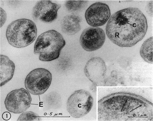
Previous serological studies have indicated that the group antigen of chlamydial organisms is composed of an acidic polysaccharide and a lipid component. The present study was undertaken in an effort to locate this polysaccharide complex by use of electron microscopy and a silver-methenamine marker. The meningopneumonitis strain of Chlamydia psittaci was propagated in HeLa-M cell culture. Organisms were purified by differential centrifugation, treatment with Genetron, and by gel filtration. After fixation and embedding, sections were obtained for electron microscopy. Sections were stained for carbohydrates with silver-methenamine. A double layer of regularly spaced silver grains of uniform size was observed at the periphery of the sectioned organisms tracing the contours of the surface membrane (cell wall). This intensity of staining was observed only when sections were oxidized with periodate prior to silver-methenamine staining. Prior treatment with 1% sodium deoxycholate resulted in a significant reduction in staining. It is considered probable that the periodate-sensitive polysaccharide found at the periphery of the organisms represents, or is a component of, the group antigen of these organisms.
以往的血清学研究表明,衣原体生物体的群抗原由一种酸性多糖和一种脂质成分组成。本研究旨在通过电子显微镜和银甲胺标记来定位这种多糖复合物。鹦鹉热衣原体的脑膜肺炎菌株在HeLa-M细胞培养物中繁殖。通过差速离心、用二氯二氟甲烷处理以及凝胶过滤对生物体进行纯化。固定和包埋后,获得用于电子显微镜检查的切片。用银甲胺对切片进行碳水化合物染色。在切片生物体的周边观察到一层由大小均匀、规则间隔的银颗粒组成的双层结构,勾勒出表面膜(细胞壁)的轮廓。只有在银甲胺染色前用高碘酸盐氧化切片时才观察到这种染色强度。用1%脱氧胆酸钠预先处理会导致染色显著减少。据认为,在生物体周边发现的对高碘酸盐敏感的多糖代表这些生物体群抗原,或者是其组成部分。