Pick E, Krejci J, Cech K, Turk J L
Immunology. 1969 Nov;17(5):741-67.
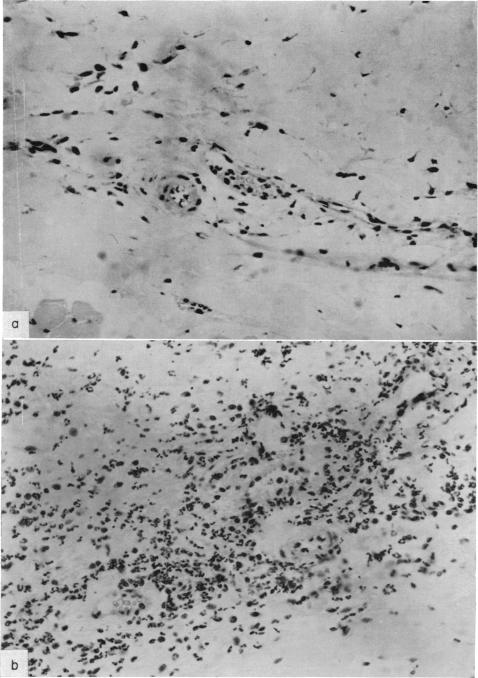
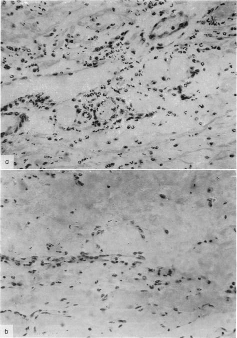
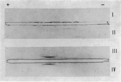
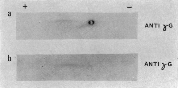
Peritoneal exudate lymphocytes (PEL), purified on glass beads and lymph node (LN) cells from guinea-pigs immunized with tubercle bacilli were cultured for 24 hours, in serum-free medium, without and with various concentrations of Tuberculin PPD. Supernatants obtained from cultures with 10 μg PPD/10 lymphocytes provoked an intense inflammatory reaction, when injected into the skin of normal guinea-pigs. PEL were more active than LN cells from the same animals. The reaction was characterized by erythema and induration, with a peak between 3 and 6 hours and histologically a mixed polymorphonuclear—mononuclear infiltrate in the dermis was seen. When fractionated on Sephadex G-200, skin activity of both PEL and LN supernatants was concentrated in a peak corresponding to the molecular weight of serum albumin, while in LN material some activity was also present in a small molecular weight peak. The active material could be separated from albumin by chromatography on DEAE-cellulose. Immunoelectrophoretic analysis of skin reactive peaks detected a slow α-globulin in both PEL and LN supernatants. PPD in the form of a complex with a protein precipitated by anti-γG antiserum, was detected in the skin-active Sephadex peak III of LN supernatants, by radioimmunoelectrophoresis. Skin activity was precipitated with ammonium sulphate at 66 per cent saturation and was destroyed by pepsin treatment. Formation of the skin-active material was depressed by Puromycin and Actinomycin-D and the development of skin inflammation was suppressed by pretreatment of the recipient with anti-lymph node extract serum. Evidence for antigen induced specific synthesis and release of an α-globulin in PEL and LN cultures was found but its relation to the skin active material is unknown.
用玻璃珠纯化的腹腔渗出淋巴细胞(PEL)以及来自用结核杆菌免疫的豚鼠的淋巴结(LN)细胞,在无血清培养基中培养24小时,分别在不添加和添加不同浓度结核菌素PPD的情况下进行培养。用10μg PPD/10个淋巴细胞培养得到的上清液,注射到正常豚鼠皮肤中时会引发强烈的炎症反应。来自同一动物的PEL比LN细胞更具活性。该反应的特征为红斑和硬结,在3至6小时达到峰值,组织学上可见真皮中有混合的多形核细胞 - 单核细胞浸润。当在Sephadex G - 200上进行分级分离时,PEL和LN上清液的皮肤活性集中在与血清白蛋白分子量相对应的峰值处,而在LN物质中,一些活性也存在于小分子质量峰值处。活性物质可通过DEAE - 纤维素柱层析与白蛋白分离。对皮肤反应性峰值进行免疫电泳分析,在PEL和LN上清液中均检测到一种慢α球蛋白。通过放射免疫电泳在LN上清液的皮肤活性Sephadex峰III中检测到与抗γG抗血清沉淀的蛋白质形成复合物形式的PPD。皮肤活性在66%饱和度的硫酸铵中沉淀,并被胃蛋白酶处理破坏。嘌呤霉素和放线菌素 - D可抑制皮肤活性物质的形成,用抗淋巴结提取物血清预处理受体可抑制皮肤炎症的发展。在PEL和LN培养物中发现了抗原诱导的α球蛋白特异性合成和释放的证据,但其与皮肤活性物质的关系尚不清楚。