Prior R B, Warner J F
Antimicrob Agents Chemother. 1974 Dec;6(6):853-5. doi: 10.1128/AAC.6.6.853.
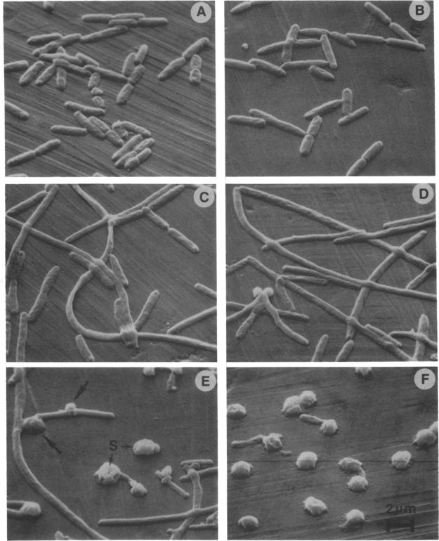
Pseudomonas aeruginosa was exposed to 0.1, 1.0, 10, 100, and 1,000 times the minimal inhibitory concentration of ticarcillin in vitro and subsequently examined with the scanning electron microscope. The morphological alterations observed were filamentation, mid-cell defects, and spheroplast formation, and these alterations were dependent upon the drug concentration.
将铜绿假单胞菌在体外暴露于替卡西林最低抑菌浓度的0.1、1.0、10、100和1000倍浓度下,随后用扫描电子显微镜进行检查。观察到的形态学改变包括丝状化、细胞中部缺陷和原生质球形成,且这些改变取决于药物浓度。