Wang N S, Huang S N, Thurlbeck W M
Am J Pathol. 1972 Jun;67(3):571-82.
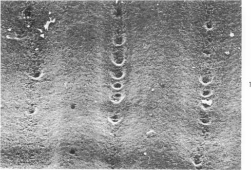
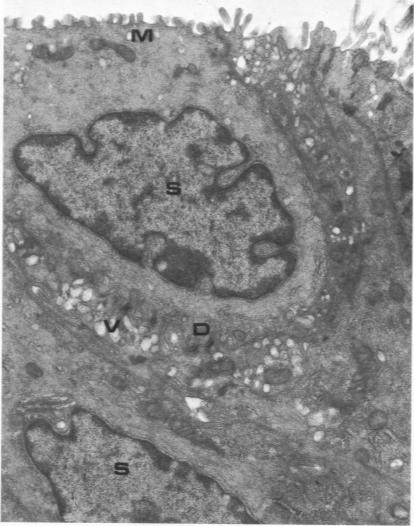
The major bronchi of swine, a dog and rabbits were examined with a scanning electron microscope and the fine structure of the openings of bronchial glands were studied three-dimensionally. The smallest areas of squamous metaplasia involved the duct openings. By examining serial sections with the light and transmission electron microscopes these early lesions were found localized at the opening of the bronchial gland duct. Cells intermediate to goblet and squamous cells were present in these lesions. Squamous metaplasia starting at this particular location is probably common and metaplasia can be an intracellular process.
用扫描电子显微镜检查了猪、狗和兔子的主支气管,并对支气管腺开口的精细结构进行了三维研究。鳞状化生面积最小的区域涉及导管开口。通过光学显微镜和透射电子显微镜检查连续切片,发现这些早期病变局限于支气管腺导管开口处。在这些病变中存在介于杯状细胞和鳞状细胞之间的细胞。从这个特定位置开始的鳞状化生可能很常见,并且化生可能是一个细胞内过程。