Long J C, Aisenberg A C, Zamecnik P C
Proc Natl Acad Sci U S A. 1974 Jun;71(6):2285-9. doi: 10.1073/pnas.71.6.2285.
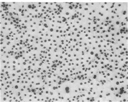
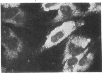
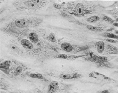
Rabbits were immunized with an antigen of specific gravity, 1.15-1.21 isolated by density gradient sedimentation of the centrifuged medium of long-term monolayer cultures derived from spleens involved by Hodgkin's disease. The globulin fraction of the antiserum was absorbed to reduce reactivity with normal cellular antigens and tissue culture components, and was tested by the indirect fluorescent antibody technique with cells from 18 different Hodgkin's disease cultures, and 16 normal cultures derived from adult spleen and fetal spleen and thymus. With anti-Hodgkin's disease globulin diluted 1:40 and 1:80, positive surface staining was observed in 48% and 41%, respectively, of viable cells from Hodgkin's disease cultures, and in less than 5% of cells cells from normal cultures. Fluorescent staining of the cytoplasm without nuclear staining was observed in 51% of acetone-fixed cells from the Hodgkin's disease cultures and in 4-8% of cells from normal cultures. Reactivity of the antiserum with Hodgkin's disease target cells could be removed by absorption of the antibody with additional antigen of density 1.15-1.21 obtained from other Hodgkin's disease cultures. Antisera to fractionated medium from a normal spleen culture and to noncultured Hodgkin's disease tumor tissue were used as controls: 2-10% of viable and acetone-fixed target cells reacted and no difference was observed between Hodgkin's disease and normal cell cultures. In vitro propagation of tumor cells from patients with Hodgkin's disease is needed for detection of the Hodgkin's disease tissue culture antigen; the antigen could not be demonstrated in noncultured Hodgkin's disease tissue.
用通过密度梯度沉降从霍奇金病累及脾脏的长期单层培养物的离心培养基中分离出的比重为1.15 - 1.21的抗原免疫兔子。吸收抗血清的球蛋白部分以降低与正常细胞抗原和组织培养成分的反应性,并通过间接荧光抗体技术用来自18种不同霍奇金病培养物以及16种来自成人脾脏、胎儿脾脏和胸腺的正常培养物的细胞进行检测。用稀释至1:40和1:80的抗霍奇金病球蛋白,分别在48%和41%的霍奇金病培养物的活细胞中观察到阳性表面染色,而在正常培养物的细胞中不到5%的细胞出现阳性表面染色。在51%的来自霍奇金病培养物的丙酮固定细胞以及4 - 8%的来自正常培养物的细胞中观察到细胞质荧光染色而无细胞核染色。抗血清与霍奇金病靶细胞的反应性可通过用从其他霍奇金病培养物获得的比重为1.15 - 1.21的额外抗原吸收抗体来消除。来自正常脾脏培养物的分级培养基的抗血清以及非培养的霍奇金病肿瘤组织的抗血清用作对照:2 - 10%的活的和丙酮固定的靶细胞有反应,并且在霍奇金病和正常细胞培养物之间未观察到差异。为了检测霍奇金病组织培养抗原,需要对霍奇金病患者的肿瘤细胞进行体外培养;在非培养的霍奇金病组织中未证实有该抗原。