• 文献检索
  • 文档翻译
  • 深度研究
  • 学术资讯
  • Suppr Zotero 插件Zotero 插件
  • 邀请有礼
  • 套餐&价格
  • 历史记录
应用&插件
Suppr Zotero 插件Zotero 插件浏览器插件Mac 客户端Windows 客户端微信小程序
定价
高级版会员购买积分包购买API积分包
服务
文献检索文档翻译深度研究API 文档MCP 服务
关于我们
关于 Suppr公司介绍联系我们用户协议隐私条款
关注我们

Suppr 超能文献

核心技术专利:CN118964589B侵权必究
粤ICP备2023148730 号-1Suppr @ 2026

文献检索

告别复杂PubMed语法,用中文像聊天一样搜索,搜遍4000万医学文献。AI智能推荐,让科研检索更轻松。

立即免费搜索

文件翻译

保留排版,准确专业,支持PDF/Word/PPT等文件格式,支持 12+语言互译。

免费翻译文档

深度研究

AI帮你快速写综述,25分钟生成高质量综述,智能提取关键信息,辅助科研写作。

立即免费体验

长期低剂量X射线对人晶状体白内障的光学及超微结构表现的影响

Influence of a prolonged period of low-dosage x-rays on the optic and ultrastructural appearances of cataract of the human lens.

作者信息

Hayes B P, Fisher R F

出版信息

Br J Ophthalmol. 1979 Jul;63(7):457-64. doi: 10.1136/bjo.63.7.457.

DOI:10.1136/bjo.63.7.457
PMID:465419
原文链接:https://pmc.ncbi.nlm.nih.gov/articles/PMC1043514/
Abstract

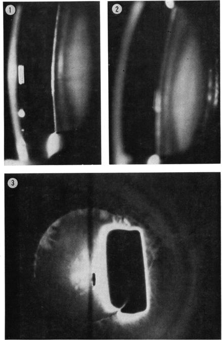
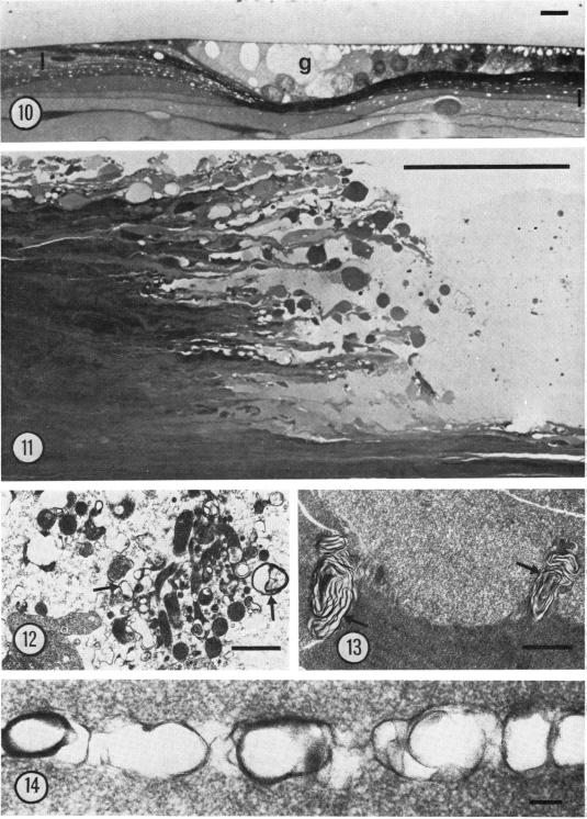
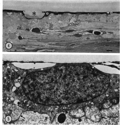
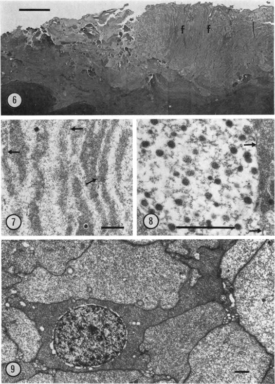
Human lenses extracted for cataract 26 years after long-term exposure to an imperfectly shielded radium source were examined by slit-lamp photography, thin-section light microscopy, and electron microscopy. Anterior epithelial cells were fibroblast-like, and germinal epithelium and vacuolated cortical fibres had accumulated at the equator. A zone of light scatter at the anterior pole corresponded to an area of breakdown of cortical lens fibres, where unusual feathery fibres were orientated perpendicular to the lens surface. Two zones of light scatter separated by a 250-microM clear interval were seen in the posterior cortex. The zone at the posterior pole corresponded to an area of fibre liquefaction and large rounded membrane whorls, while the deeper zone comprised small flattened membrane whorls. The characteristic plaques of swollen abnormal cells described in previous histological studies of x-ray cataract were not present. This and other differences probably reflect the extremely long time course and repeated subliminal doses to which the patient was exposed.

摘要

对长期暴露于防护不完善的镭源26年后因白内障而摘除的人晶状体进行了裂隙灯摄影、薄切片光学显微镜检查和电子显微镜检查。前上皮细胞呈成纤维细胞样,生发上皮和空泡化的皮质纤维在赤道处积聚。前极的光散射区对应于皮质晶状体纤维破裂的区域,此处异常的羽毛状纤维垂直于晶状体表面排列。在后皮质中可见两个由250微米透明间隔隔开的光散射区。后极的区域对应于纤维液化和大的圆形膜涡旋的区域,而较深的区域由小的扁平膜涡旋组成。先前关于X射线白内障的组织学研究中描述的肿胀异常细胞的特征性斑块不存在。这种差异以及其他差异可能反映了患者暴露于其中的极长时间过程和反复的阈下剂量。

https://cdn.ncbi.nlm.nih.gov/pmc/blobs/e357/1043514/f98ba7de2e9e/brjopthal00211-0011-a.jpg
https://cdn.ncbi.nlm.nih.gov/pmc/blobs/e357/1043514/5be1ee26e473/brjopthal00211-0007-a.jpg
https://cdn.ncbi.nlm.nih.gov/pmc/blobs/e357/1043514/b58b94df7306/brjopthal00211-0008-a.jpg
https://cdn.ncbi.nlm.nih.gov/pmc/blobs/e357/1043514/a452ddd40dc4/brjopthal00211-0009-a.jpg
https://cdn.ncbi.nlm.nih.gov/pmc/blobs/e357/1043514/f98ba7de2e9e/brjopthal00211-0011-a.jpg

相似文献

1
Influence of a prolonged period of low-dosage x-rays on the optic and ultrastructural appearances of cataract of the human lens.长期低剂量X射线对人晶状体白内障的光学及超微结构表现的影响
Br J Ophthalmol. 1979 Jul;63(7):457-64. doi: 10.1136/bjo.63.7.457.
2
Ultrastructural appearances of a lens with marked polychromatic lustre: evidence for diffraction as a cause.具有显著多色光泽的晶状体的超微结构外观:衍射作为病因的证据。
Br J Ophthalmol. 1984 Dec;68(12):850-8. doi: 10.1136/bjo.68.12.850.
3
Ultrastructure of UVR-B-induced cataract and repair visualized with electron microscopy.用电子显微镜观察UVR-B诱导的白内障及其修复的超微结构。
Acta Ophthalmol. 2014 Nov;92(7):635-43. doi: 10.1111/aos.12376. Epub 2014 Mar 25.
4
Development and repair of cataract induced by ultraviolet radiation.紫外线诱导白内障的发生与修复
Ophthalmic Res. 2000;32 Suppl 1:ii-iii; 1-44.
5
[A case of radiation cataract resulting from accidental irradiation of radioisotope].[一例放射性核素意外照射导致的放射性白内障]
Nippon Ganka Gakkai Zasshi. 1993 Mar;97(3):419-25.
6
Evolution of damage in the lens after in vivo close to threshold exposure to UV-B radiation: cytomorphological study of apoptosis.活体近阈值紫外线 B 辐射后晶状体损伤的演变:细胞形态学研究细胞凋亡。
Exp Eye Res. 2010 Sep;91(3):369-77. doi: 10.1016/j.exer.2010.06.009. Epub 2010 Jun 17.
7
Ultrastructural lens changes in X-ray induced cataract of the rat.大鼠X射线诱导性白内障的超微结构晶状体变化
Acta Ophthalmol (Copenh). 1978;56(4):587-98. doi: 10.1111/j.1755-3768.1978.tb01371.x.
8
A histological study on the cataractogenic effects of heavy charged particles.重带电粒子致白内障作用的组织学研究
Proc Natl Sci Counc Repub China B. 1987 Jan;11(1):18-28.
9
Histopathological study of hereditary cataractous lenses in SCR strain rat.SCR品系大鼠遗传性白内障晶状体的组织病理学研究
Exp Eye Res. 1993 Nov;57(5):567-76. doi: 10.1006/exer.1993.1161.
10
Repair in the rat lens after threshold ultraviolet radiation injury.阈值紫外线辐射损伤后大鼠晶状体的修复
Invest Ophthalmol Vis Sci. 2000 Jan;41(1):204-12.

引用本文的文献

1
Effect of Instrument Navigation on C-arm Radiation and Time during Spinal Procedures: A Clinical Evaluation.仪器导航对脊柱手术中C型臂辐射及手术时间的影响:一项临床评估
Int J Spine Surg. 2020 Jun 30;14(3):375-381. doi: 10.14444/7049. eCollection 2020 Jun.
2
Lauriston S. Taylor Lecture on radiation protection and measurements: what makes particle radiation so effective?劳里斯顿·S·泰勒辐射防护与测量讲座:是什么让粒子辐射如此有效?
Health Phys. 2012 Nov;103(5):508-28. doi: 10.1097/HP.0b013e31826a5b85.
3
A clinical study of radiation cataract formation in adult life following gamma irradiation of the lens in early childhood.

本文引用的文献

1
Clinical and pathological characteristics of radiation cataract.放射性白内障的临床及病理特征
AMA Arch Ophthalmol. 1952 Jan;47(1):55-70. doi: 10.1001/archopht.1952.01700030058006.
2
Experimental radiation cataracts. I. Cataracts in the rabbit following single x-ray exposure.实验性辐射性白内障。I. 单次X射线照射后家兔的白内障
AMA Arch Ophthalmol. 1951 May;45(5):508-22.
3
[ULTRASTRUCTURAL CHANGES OF THE CRYSTALLINE LENS IN CERTAIN EXPERIMENTAL AND HUMAN CATARACTS].[某些实验性和人类白内障中晶状体的超微结构变化]
一项关于儿童早期晶状体接受伽马射线照射后成年期放射性白内障形成的临床研究。
Br J Ophthalmol. 1997 Apr;81(4):261-6. doi: 10.1136/bjo.81.4.261.
4
Ultrastructure and elastic changes of regenerated basement membrane in normal and diabetic rats.正常及糖尿病大鼠再生基底膜的超微结构与弹性变化
Br J Exp Pathol. 1982 Jun;63(3):341-50.
5
Ultrastructural appearances of a lens with marked polychromatic lustre: evidence for diffraction as a cause.具有显著多色光泽的晶状体的超微结构外观:衍射作为病因的证据。
Br J Ophthalmol. 1984 Dec;68(12):850-8. doi: 10.1136/bjo.68.12.850.
6
Possible cataractogenic effect of radionuclides deposited within the eye from the blood stream.放射性核素从血流中沉积在眼内可能产生的致白内障作用。
Br J Ophthalmol. 1985 Mar;69(3):219-27. doi: 10.1136/bjo.69.3.219.
Bull Mem Soc Fr Ophtalmol. 1963;76:193-208.
4
The use of lead citrate at high pH as an electron-opaque stain in electron microscopy.在电子显微镜检查中,将高pH值的柠檬酸铅用作电子不透明染色剂。
J Cell Biol. 1963 Apr;17(1):208-12. doi: 10.1083/jcb.17.1.208.
5
Junctions between intimately apposed cell membranes in the vertebrate brain.脊椎动物大脑中紧密相邻细胞膜之间的连接。
J Cell Biol. 1969 Mar;40(3):648-77. doi: 10.1083/jcb.40.3.648.
6
Senile cataract. A comparative study between lens fibre stress and cuneiform opacity formation.
Trans Ophthalmol Soc U K (1962). 1970;90:93-109.
7
[On the atomic bomb radiation cataract].[论原子弹辐射性白内障]
Nippon Ganka Gakkai Zasshi. 1973;77(3):305-9.
8
Changes in the lens related to the reduction of transparency.晶状体透明度降低相关的变化。
Exp Eye Res. 1973 Jun;16(1):29-39. doi: 10.1016/0014-4835(73)90234-0.
9
Senile changes in the fine structure of the lens in the rudd(Scardinius eryopthalmicus).赤睛鱼(Scardinius eryopthalmicus)晶状体精细结构的衰老变化。
Exp Eye Res. 1974 Jun;18(6):571-7. doi: 10.1016/0014-4835(74)90063-3.
10
Changes in lens fibres after damage to the lens capsule.晶状体囊受损后晶状体纤维的变化。
Trans Ophthalmol Soc U K (1962). 1976 Jul;96(2):278-84.