Ashraf M
Am J Pathol. 1979 Nov;97(2):411-32.
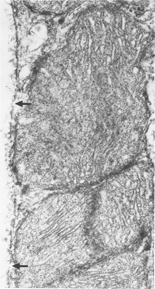
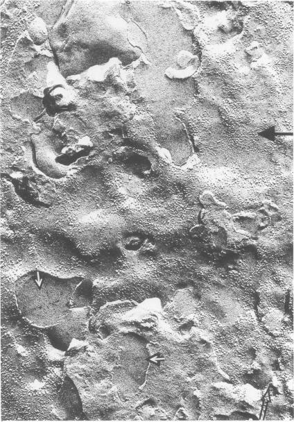
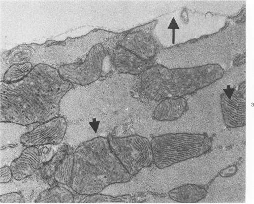
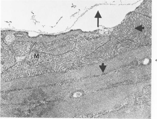
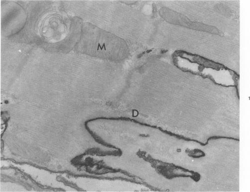
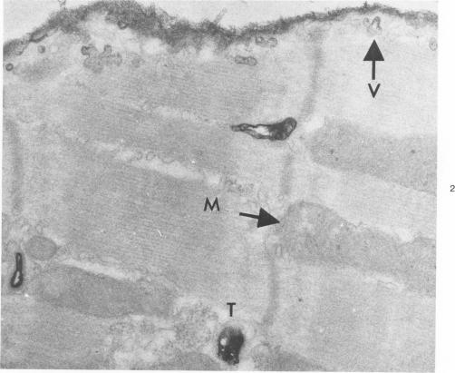
Effects of calcium-free perfusion and calcium-free perfusion followed by reperfusion with calcium on sarcolemmal structure, sarcolemmal permeability, and creatine phosphokinase loss were investigated in isolated perfused rat hearts. Release of creatine phosphokinase was significant (P less than 0.0002) after 4-5 minutes of perfusion with Ca++-free medium, but later releases in comparison to their immediately preceding periods became significant only after more than 20-minute perfusion. Poor correlation between enzyme loss and lanthanum permeability prior to 20 minutes of Ca++-free perfusion was noted. After 20 minutes of Ca++-free perfusion, the basal lamina was separated from the plasma membrane, and lanthanum was seen in the cytoplasm. The intramembranous particles began to aggregate at that time. The morphologic and enzymatic changes were dramatic following reperfusion of calcium-free perfused hearts. Morphologic changes in these hearts included separation of basal lamina, cellular separation at the intercalated disk, dissolution of actin filaments at the region of I band, contraction bands, cell swelling, and staining or filling of mitochondrial membranes with La+++. Increased sarcolemmal permeability was associated with tears and aggregation of intramembranous particles in the sarcolemmal lipid bilayers. These results suggest that reperfusion of Ca++-free perfused cells causes irreversible damage to the sarcolemmal lipid bilayer, and the degree of alterations induced in the cells is dependent upon the initial duration of Ca++-free perfusion.
在离体灌注大鼠心脏中,研究了无钙灌注以及无钙灌注后再用钙复灌对肌膜结构、肌膜通透性和肌酸磷酸激酶损失的影响。在用无Ca++培养基灌注4 - 5分钟后,肌酸磷酸激酶的释放显著(P小于0.0002),但与紧接的前一时期相比,只有在灌注超过20分钟后,后续的释放才变得显著。在无Ca++灌注20分钟之前,酶损失与镧通透性之间的相关性较差。无Ca++灌注20分钟后,基膜与质膜分离,在细胞质中可见镧。此时膜内颗粒开始聚集。无钙灌注心脏再灌注后,形态学和酶学变化显著。这些心脏的形态学变化包括基膜分离、闰盘处细胞分离、I带区域肌动蛋白丝溶解、收缩带、细胞肿胀以及线粒体膜被La+++染色或填充。肌膜通透性增加与肌膜脂质双层中膜内颗粒的撕裂和聚集有关。这些结果表明,无Ca++灌注细胞的再灌注会对肌膜脂质双层造成不可逆损伤,细胞中诱导的改变程度取决于无Ca++灌注的初始持续时间。