• 文献检索
  • 文档翻译
  • 深度研究
  • 学术资讯
  • Suppr Zotero 插件Zotero 插件
  • 邀请有礼
  • 套餐&价格
  • 历史记录
应用&插件
Suppr Zotero 插件Zotero 插件浏览器插件Mac 客户端Windows 客户端微信小程序
定价
高级版会员购买积分包购买API积分包
服务
文献检索文档翻译深度研究API 文档MCP 服务
关于我们
关于 Suppr公司介绍联系我们用户协议隐私条款
关注我们

Suppr 超能文献

核心技术专利:CN118964589B侵权必究
粤ICP备2023148730 号-1Suppr @ 2026

文献检索

告别复杂PubMed语法,用中文像聊天一样搜索,搜遍4000万医学文献。AI智能推荐,让科研检索更轻松。

立即免费搜索

文件翻译

保留排版,准确专业,支持PDF/Word/PPT等文件格式,支持 12+语言互译。

免费翻译文档

深度研究

AI帮你快速写综述,25分钟生成高质量综述,智能提取关键信息,辅助科研写作。

立即免费体验

珍珠鸡(Numida meleagris)睾丸输出管的结构。

The structure of the excurrent ducts of the testis of the guinea-fowl (Numida meleagris).

作者信息

Aire T A, Ayeni J S, Olowo-Okorun M O

出版信息

J Anat. 1979 Oct;129(Pt 3):633-43.

PMID:541246
原文链接:https://pmc.ncbi.nlm.nih.gov/articles/PMC1233029/
Abstract

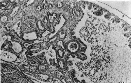
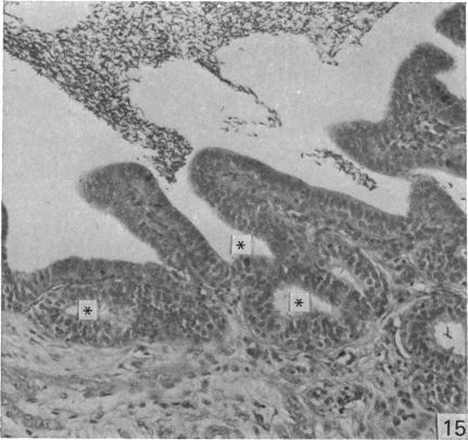
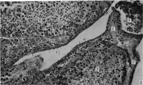
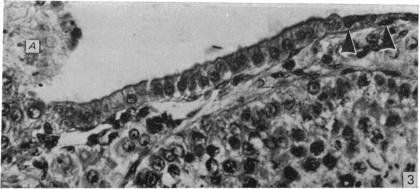
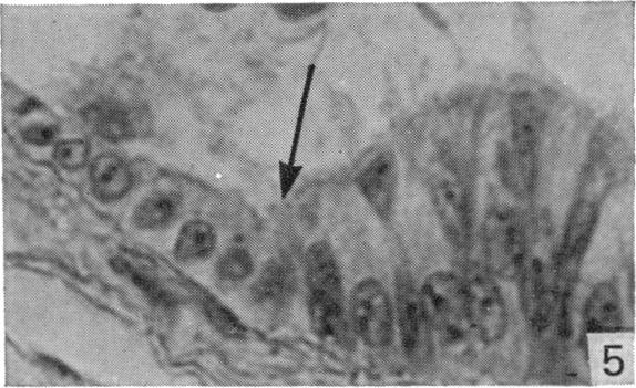
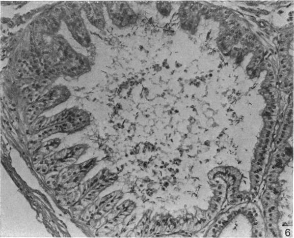
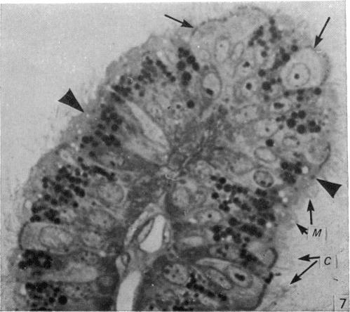
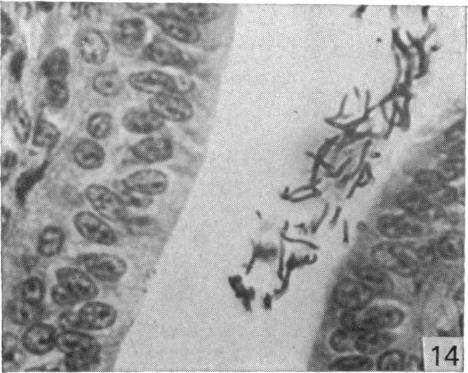
The epididymal region of the guinea-fowl was studied in sexually mature birds. The structure of the epididymal region was generally similar to that already described for the domestic fowl, turkey and Japanese quail. Well formed, intratesticular tubuli recti was seen connecting the seminiferous tubules with the rete testis. The latter consists of both intracapsular and extracapsular portions. Six main cell types were recognised in the region: the rete testis was lined by squamous cells, the proximal efferent ductules by ciliated and non-ciliated Type I cells, the distal efferent ductule by ciliated and non-ciliated Type II cells, and the connecting ductules, ductus epididymidis and ductus deferens were lined by non-ciliated Type III and basal cells. The cell classification adopted in this study is discussed.

摘要

对性成熟珍珠鸡的附睾区域进行了研究。附睾区域的结构总体上与已描述的家鸡、火鸡和日本鹌鹑的结构相似。可见发育良好的睾丸内直小管将生精小管与睾丸网相连。睾丸网由囊内和囊外部分组成。该区域识别出六种主要细胞类型:睾丸网内衬扁平细胞,近端输出小管内衬纤毛和非纤毛I型细胞,远端输出小管内衬纤毛和非纤毛II型细胞,连接小管、附睾管和输精管内衬非纤毛III型细胞和基底细胞。讨论了本研究采用的细胞分类。

https://cdn.ncbi.nlm.nih.gov/pmc/blobs/bdfd/1233029/02bf6bd191a6/janat00239-0188-b.jpg
https://cdn.ncbi.nlm.nih.gov/pmc/blobs/bdfd/1233029/1ffdeb4ce9f0/janat00239-0181-a.jpg
https://cdn.ncbi.nlm.nih.gov/pmc/blobs/bdfd/1233029/a0bee3dd7d2a/janat00239-0181-b.jpg
https://cdn.ncbi.nlm.nih.gov/pmc/blobs/bdfd/1233029/b255179bb452/janat00239-0182-a.jpg
https://cdn.ncbi.nlm.nih.gov/pmc/blobs/bdfd/1233029/43f5a0f7e833/janat00239-0182-b.jpg
https://cdn.ncbi.nlm.nih.gov/pmc/blobs/bdfd/1233029/da4ab0eb30ef/janat00239-0183-a.jpg
https://cdn.ncbi.nlm.nih.gov/pmc/blobs/bdfd/1233029/ed814153d5c4/janat00239-0183-b.jpg
https://cdn.ncbi.nlm.nih.gov/pmc/blobs/bdfd/1233029/42198532cab5/janat00239-0184-a.jpg
https://cdn.ncbi.nlm.nih.gov/pmc/blobs/bdfd/1233029/bb90406e5eb0/janat00239-0185-a.jpg
https://cdn.ncbi.nlm.nih.gov/pmc/blobs/bdfd/1233029/1813cff90156/janat00239-0185-b.jpg
https://cdn.ncbi.nlm.nih.gov/pmc/blobs/bdfd/1233029/607ae1c7a8bd/janat00239-0186-a.jpg
https://cdn.ncbi.nlm.nih.gov/pmc/blobs/bdfd/1233029/e44f8574081c/janat00239-0186-b.jpg
https://cdn.ncbi.nlm.nih.gov/pmc/blobs/bdfd/1233029/89c7aefdf1c3/janat00239-0187-a.jpg
https://cdn.ncbi.nlm.nih.gov/pmc/blobs/bdfd/1233029/363bb8b49056/janat00239-0187-b.jpg
https://cdn.ncbi.nlm.nih.gov/pmc/blobs/bdfd/1233029/699fc19c0958/janat00239-0188-a.jpg
https://cdn.ncbi.nlm.nih.gov/pmc/blobs/bdfd/1233029/02bf6bd191a6/janat00239-0188-b.jpg

相似文献

1
The structure of the excurrent ducts of the testis of the guinea-fowl (Numida meleagris).珍珠鸡(Numida meleagris)睾丸输出管的结构。
J Anat. 1979 Oct;129(Pt 3):633-43.
2
The epididymal region of the Japanese quail (Coturnix coturnix japonica).日本鹌鹑(Coturnix coturnix japonica)的附睾区域。
Acta Anat (Basel). 1979;103(3):305-12.
3
Morphology of the epididymal region and ductus deferens of the turkey (Meleagris gallopavo).火鸡(吐绶鸡)附睾区域和输精管的形态学
J Anat. 1976 Nov;122(Pt 2):241-52.
4
Immunohistochemistry of the cytoskeleton in the excurrent ducts of the testis in birds of the Galloanserae monophyly.鸡雁小纲鸟类睾丸输出小管细胞骨架的免疫组织化学研究
Cell Tissue Res. 2008 Aug;333(2):311-21. doi: 10.1007/s00441-008-0631-7. Epub 2008 Jun 19.
5
The ductuli efferentes of the epididymal region of birds.鸟类附睾区域的输出小管。
J Anat. 1980 Jun;130(Pt 4):707-23.
6
Micro-stereological study of the avian epididymal region.鸟类附睾区域的微观体视学研究。
J Anat. 1979 Dec;129(Pt 4):703-6.
7
Ultrastructural study of the luminal surface of the ducts of the epididymis of gallinaceous birds.
Onderstepoort J Vet Res. 2000 Sep;67(3):191-9.
8
Observations on the anterior testicular ducts in snakes with emphasis on sea snakes and ultrastructure in the yellow-bellied sea snake, Pelamis platurus.对蛇类睾丸前导管的观察,重点是海蛇以及黄腹海蛇(长吻海蛇)的超微结构。
J Morphol. 2012 Mar;273(3):324-36. doi: 10.1002/jmor.11025. Epub 2011 Oct 24.
9
Morphological changes in the efferent ducts during the main phases of the reproductive cycle of birds.
J Morphol. 2002 Jul;253(1):64-75. doi: 10.1002/jmor.1113.
10
Immunolocalization of aquaporins 1, 2 and 7 in rete testis, efferent ducts, epididymis and vas deferens of adult dog.水通道蛋白1、2和7在成年犬睾丸网、输出小管、附睾和输精管中的免疫定位
Cell Tissue Res. 2008 May;332(2):329-35. doi: 10.1007/s00441-008-0592-x. Epub 2008 Mar 14.

引用本文的文献

1
Photoperiodic-dependent histomorphological changes in the excurrent duct system of helmeted guinea fowl subjected to short day (8L:16D), long-day (16L:8D) light/dark cycles and exogenous melatonin.接受短日照(8小时光照:16小时黑暗)、长日照(16小时光照:8小时黑暗)光暗循环及外源性褪黑素处理的盔珠鸡输出管系统中光周期依赖性组织形态学变化
Vet Anim Sci. 2022 Dec 22;19:100282. doi: 10.1016/j.vas.2022.100282. eCollection 2023 Mar.
2
Histological structure and age-related changes in the luminal diameter of the excurrent duct system of guinea cocks and associated changes in testosterone concentrations.豚鸡输出管系统的组织学结构、管腔直径的年龄相关变化以及睾酮浓度的相关变化。
Can J Vet Res. 2018 Jan;82(1):60-65.
3

本文引用的文献

1
An electron microscopic study of the ductuli efferentes and rete testis of the guinea pig.豚鼠输出小管和睾丸网的电子显微镜研究。
J Biophys Biochem Cytol. 1958 Mar 25;4(2):219-26. doi: 10.1083/jcb.4.2.219.
2
The male reproductive tract of the fowl.家禽的雄性生殖道。
J Anat. 1957 Jan;91(1):116-29.
3
[Constitution of the epididymis of the rooster].[公鸡附睾的结构]
The ductuli efferentes of the epididymal region of birds.
鸟类附睾区域的输出小管。
J Anat. 1980 Jun;130(Pt 4):707-23.
4
The epididymis and its development in ratite birds (ostrich, emu, rhea).平胸鸟类(鸵鸟、鸸鹋、美洲鸵)附睾及其发育
Anat Embryol (Berl). 1981;162(3):281-99. doi: 10.1007/BF00299973.
5
Surface morphology of the ducts of the epididymal region of the drake (Anas platyrhynchos) as revealed by scanning and transmission electron microscopy.通过扫描电子显微镜和透射电子显微镜揭示的公鸭(绿头鸭)附睾区域导管的表面形态。
J Anat. 1982 Oct;135(Pt 3):513-20.
6
The rete testis of birds.鸟类的睾丸网
J Anat. 1982 Aug;135(Pt 1):97-110.
7
A study of the rete testis epithelium in several wild birds.对几种野生鸟类睾丸网上皮的一项研究。
J Anat. 1984 Jan;138 ( Pt 1)(Pt 1):139-52.
8
The ultrastructure of the epithelium of the ductuli efferentes testis in the common starling (Sturnus vulgaris).家八哥(Sturnus vulgaris)睾丸输出小管上皮的超微结构。
J Anat. 1985 Mar;140 ( Pt 2)(Pt 2):189-203.
9
Studies of fluid and spermatozoal transport in the extratesticular genital ducts of the Japanese quail.日本鹌鹑睾丸外生殖管道中液体和精子运输的研究。
J Anat. 1988 Apr;157:1-11.
C R Seances Soc Biol Fil. 1955 Apr;149(7-8):687-9.
4
On the structure of the epididymal region and ductus deferens of the domestic fowl (Gallus domesticus).关于家鸡(原鸡)附睾区域和输精管的结构
J Anat. 1971 Sep;109(Pt 3):423-35.
5
The fine structure of the epithelial lining of the ex-current duct system of the testis of the domestic fowl (Gallus domesticus).
Q J Exp Physiol Cogn Med Sci. 1972 Jul;57(3):271-95. doi: 10.1113/expphysiol.1972.sp002162.
6
Morphology of the epididymis of the cock (Gallus domesticus) and its effect upon the steroid sex hormone synthesis. I. Ontogenesis, morphology and distribution of the epididymis.公鸡(家鸡)附睾的形态及其对类固醇性激素合成的影响。I. 附睾的个体发生、形态及分布
Anat Embryol (Berl). 1975 Dec 23;148(2):175-96. doi: 10.1007/BF00315268.
7
Morphology of the epididymal region and ductus deferens of the turkey (Meleagris gallopavo).火鸡(吐绶鸡)附睾区域和输精管的形态学
J Anat. 1976 Nov;122(Pt 2):241-52.
8
Ultrastructure of epithelial cells in the epididymal region of the turkey (Meleagris gallopavo).火鸡(吐绶鸡)附睾区域上皮细胞的超微结构
J Anat. 1977 Dec;124(Pt 3):765-78.
9
The epididymal region of the Japanese quail (Coturnix coturnix japonica).日本鹌鹑(Coturnix coturnix japonica)的附睾区域。
Acta Anat (Basel). 1979;103(3):305-12.