Aire T A
J Anat. 1982 Oct;135(Pt 3):513-20.
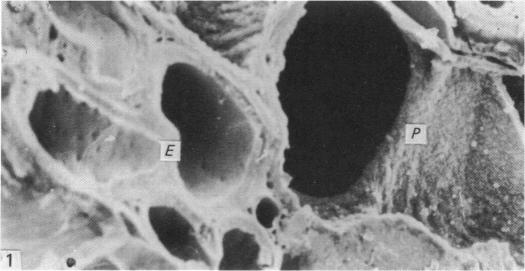
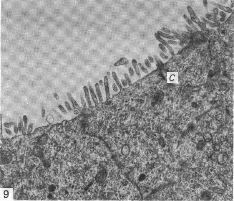
Both scanning (SEM) and transmission (TEM) electron microscopic studies of the major ductules and ducts of the perfused epididymal region of the drake were reported. The SEM correlated with TEM studies and confirmed some previous observations that the non-ciliated Types I and II cells in the proximal and distal efferent ductules, respectively, possessed apical microvilli as distinct from the cilia of the ciliated cells. The relative number of each cell type in each duct was also revealed. All microvilli and cilia were regular in shape. The connecting and epididymal ducts showed 'craters' scattered over their entire epithelial surfaces. Also, a single cilium projected from most of the cells of the epithelial lining into the lumen of these ducts. The name, 'uniciliated cell' has been suggested to describe this cell which has, until now, been referred to as the non-ciliated Type III cell (Aire, 1980; Aire et al. 1979). Neither bulbous microvilli nor blebbing of the apical plasmalemma of the cells occurred in properly fixed tissues.
本文报道了对公鸭灌注附睾区域的主要小导管和导管进行扫描电子显微镜(SEM)和透射电子显微镜(TEM)研究的结果。扫描电子显微镜研究与透射电子显微镜研究结果相关,并证实了先前的一些观察结果,即近端和远端输出小管中分别的非纤毛I型和II型细胞具有顶端微绒毛,这与纤毛细胞的纤毛不同。还揭示了每个导管中每种细胞类型的相对数量。所有微绒毛和纤毛形状规则。连接管和附睾管在其整个上皮表面散布着“火山口”。此外,上皮衬里的大多数细胞有一根纤毛伸入这些导管的管腔。有人建议用“单纤毛细胞”这个名称来描述这种细胞,到目前为止它一直被称为非纤毛III型细胞(艾尔,1980年;艾尔等人,1979年)。在固定良好的组织中,细胞顶端质膜既没有出现球状微绒毛,也没有出现起泡现象。