Virgilio R, González C, Muñoz N, Mendoza S
J Bacteriol. 1966 May;91(5):2018-24. doi: 10.1128/jb.91.5.2018-2024.1966.
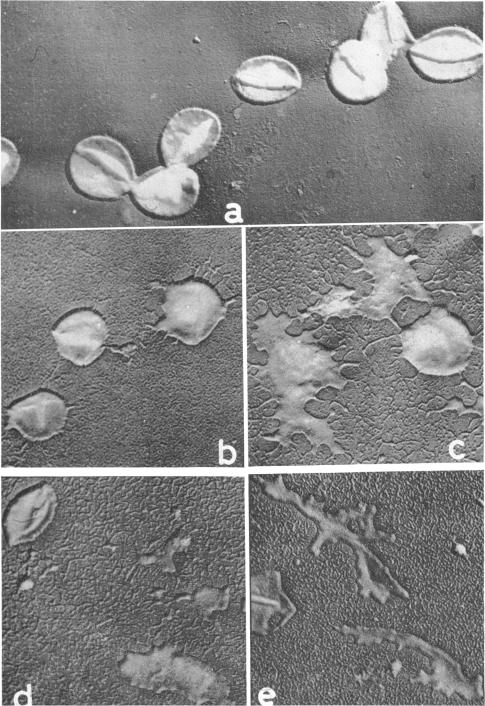
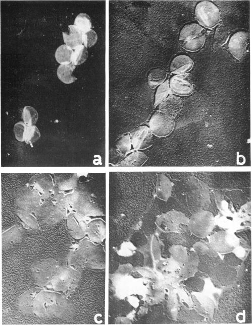
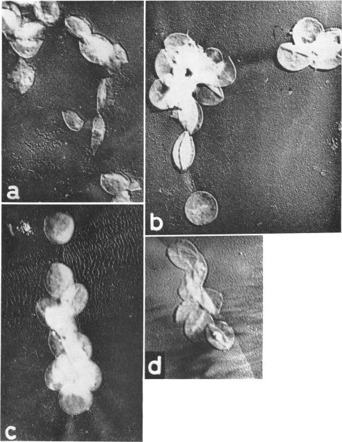
Virgilio, Rafael (Escuela de Química y Farmacia, Universidad de Chile, Santiago, Chile), C. González, Nubia Muñoz, and Silvia Mendoza. Electron microscopy of Staphylococcus aureus cell wall lysis. J. Bacteriol. 91:2018-2024. 1966.-A crude suspension of Staphylococcus aureus cell walls (strain Cowan III) in buffer solution was shown by electron microscopy to lyse slightly after 16 hr, probably owing to the action of autolysin. The lysis was considerably faster and more intense after the addition of lysozyme. A remarkable reduction in thickness and rigidity of the cell walls, together with the appearance of many irregular protrusions in their outlines, was observed after 2 hr; after 16 hr, there remained only a few recognizable cell wall fragments but many residual particulate remnants. When autolysin was previously inactivated by trypsin, there was a complete inhibition of the lytic action of lysozyme; on the other hand, when autolysin was inactivated by heat and lysozyme was added, a distinct decrease in the thickness of the cell walls was observed, but there was no destruction of the walls. The lytic action of lysozyme, after treatment with hot 5% trichloroacetic acid, gave rise to a marked dissolution of the structure of the cell walls, which became lost against the background, without, however, showing ostensible alteration of wall outlines. From a morphological point of view, the lytic action of autolysin plus lysozyme was quite different from that of trichloroacetic acid plus lysozyme, as shown by electron micrographs, but in both cases it was very intense. This would suggest different mechanisms of action for these agents.
维吉尔,拉斐尔(智利圣地亚哥智利大学化学与药学院),C. 冈萨雷斯、努维娅·穆尼奥斯和西尔维娅·门多萨。金黄色葡萄球菌细胞壁裂解的电子显微镜观察。《细菌学杂志》91:2018 - 2024。1966年。——通过电子显微镜观察发现,金黄色葡萄球菌(考恩III菌株)细胞壁在缓冲溶液中的粗悬液在16小时后略有裂解,可能是由于自溶素的作用。加入溶菌酶后,裂解速度明显加快且更为剧烈。2小时后观察到细胞壁厚度和刚性显著降低,其轮廓出现许多不规则突起;16小时后,仅剩下少数可识别的细胞壁碎片,但有许多残留的颗粒残余物。当自溶素预先被胰蛋白酶灭活时,溶菌酶的裂解作用完全受到抑制;另一方面,当自溶素被加热灭活并加入溶菌酶时,观察到细胞壁厚度明显降低,但细胞壁并未被破坏。用热的5%三氯乙酸处理后,溶菌酶的裂解作用导致细胞壁结构明显溶解,在背景中消失,然而,细胞壁轮廓并未出现明显改变。从形态学角度来看,如电子显微照片所示,自溶素加溶菌酶的裂解作用与三氯乙酸加溶菌酶的裂解作用有很大不同,但在两种情况下裂解作用都非常强烈。这表明这些试剂的作用机制不同。