Ringvold A, Davanger M
Br J Ophthalmol. 1981 Feb;65(2):138-41. doi: 10.1136/bjo.65.2.138.
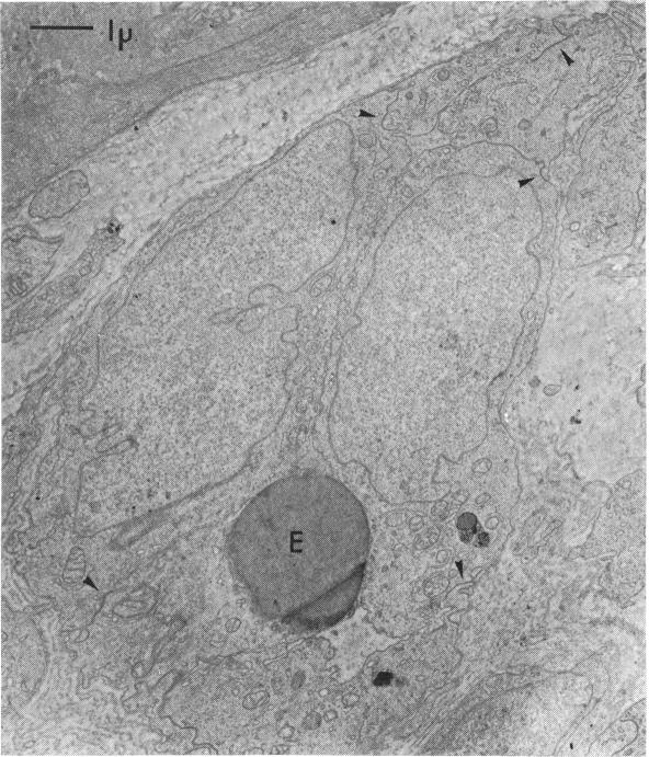
The iris vascular bed of eyes with pseudoexfoliation syndrome has been studied with electron microscopy. Two of the eyes suffered from painful glaucoma, whereas the third showed normal intraocular pressure. The following changes were observed: (1) deposits of pseudoexfoliation material lying adjacent to the endothelial wall; (2) thin vessel basement membrane, sometimes even interrupted; (3) extreme reduction of vessel lumina through increased volume of the endothelial cells; (4) fenestration of the endothelial wall. It is suggested that the iris neovascularisation in eyes with pseudoexfoliation syndrome is due to an obstruction of iris vessels causing tissue hypoxia.
已采用电子显微镜对假性剥脱综合征患者眼睛的虹膜血管床进行了研究。其中两只眼睛患有疼痛性青光眼,而第三只眼睛眼压正常。观察到以下变化:(1)假性剥脱物质沉积于内皮细胞壁附近;(2)血管基底膜变薄,有时甚至中断;(3)内皮细胞体积增加导致血管腔极度狭窄;(4)内皮细胞壁出现窗孔。有人提出,假性剥脱综合征患者眼睛的虹膜新生血管形成是由于虹膜血管阻塞导致组织缺氧所致。