Meades K V, Francis I C, Kappagoda M B, Filipic M
Br J Ophthalmol. 1986 Apr;70(4):290-4. doi: 10.1136/bjo.70.4.290.
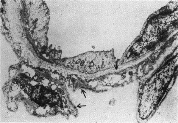
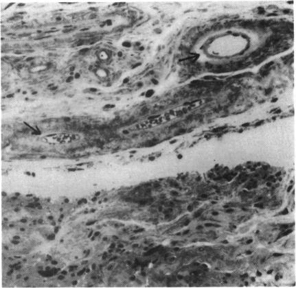
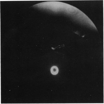
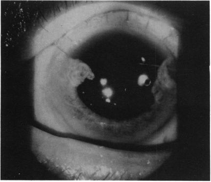
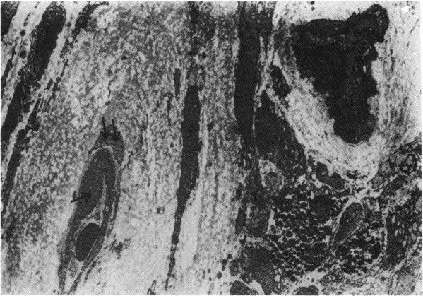
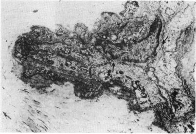
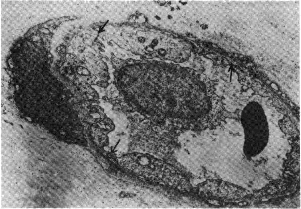
A patient who had been observed to have an iris microhaemangioma (capillary haemangioma), confirmed on fluorescein iris angiography, came to cataract surgery. The lesion was excised at the time of surgery and submitted to light and electron microscopic study. It had the features of a hamartoma of the capillary haemangioma type, with its characteristics being specific for vessels seen in iris tissue.
一名经荧光素虹膜血管造影确诊患有虹膜微血管瘤(毛细血管血管瘤)的患者前来接受白内障手术。病变在手术时被切除,并进行了光镜和电镜研究。它具有毛细血管血管瘤型错构瘤的特征,其特点是虹膜组织中所见血管特有的。