• 文献检索
  • 文档翻译
  • 深度研究
  • 学术资讯
  • Suppr Zotero 插件Zotero 插件
  • 邀请有礼
  • 套餐&价格
  • 历史记录
应用&插件
Suppr Zotero 插件Zotero 插件浏览器插件Mac 客户端Windows 客户端微信小程序
定价
高级版会员购买积分包购买API积分包
服务
文献检索文档翻译深度研究API 文档MCP 服务
关于我们
关于 Suppr公司介绍联系我们用户协议隐私条款
关注我们

Suppr 超能文献

核心技术专利:CN118964589B侵权必究
粤ICP备2023148730 号-1Suppr @ 2026

文献检索

告别复杂PubMed语法,用中文像聊天一样搜索,搜遍4000万医学文献。AI智能推荐,让科研检索更轻松。

立即免费搜索

文件翻译

保留排版,准确专业,支持PDF/Word/PPT等文件格式,支持 12+语言互译。

免费翻译文档

深度研究

AI帮你快速写综述,25分钟生成高质量综述,智能提取关键信息,辅助科研写作。

立即免费体验

黄色粘球菌发育过程中的碘化作用。

Iodination of Myxococcus xanthus during development.

作者信息

Maeba P Y

出版信息

J Bacteriol. 1983 Sep;155(3):1033-41. doi: 10.1128/jb.155.3.1033-1041.1983.

DOI:10.1128/jb.155.3.1033-1041.1983
PMID:6411682
原文链接:https://pmc.ncbi.nlm.nih.gov/articles/PMC217796/
Abstract

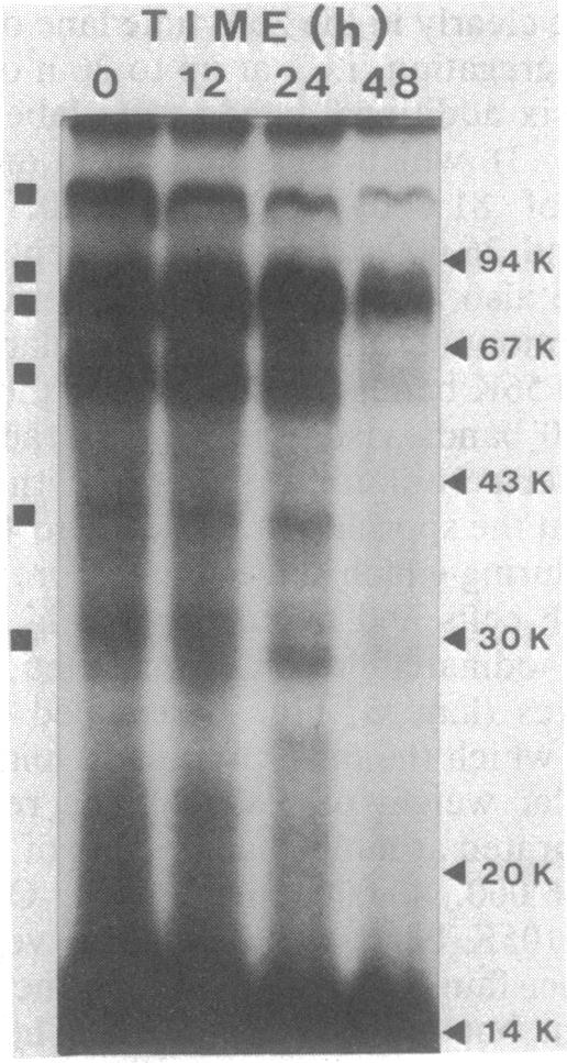
Intact cells of Myxococcus xanthus were iodinated with [125I]lactoperoxidase to permit examination of the surface components accessible to labeling during cell development. Vegetative cells, starved on a defined solid medium, aggregated, formed fruiting bodies, and produced myxospores. Cells collected at different stages were iodinated, and their proteins were analyzed by one- and two-dimensional electrophoresis and autoradiography. One-dimensional electrophoresis revealed six iodinated bands in vegetative cell extracts. During development, 10 radioactive bands were detected, 4 of which migrated to the same positions as those of vegetative cells. Only six bands were detected in purified, labeled myxospores. Of these, one band possessed mobility similar to that of labeled vegetative cell proteins, whereas the other bands possessed mobility similar to that detected in developing cells. Analysis of two-dimensional gels indicated that at least 14 proteins were iodinated in vegetative cells, one of which was intensely labeled (protein b). Another of the proteins (protein a) was labeled throughout development. During development, about 30 proteins were iodinated and the prominently labeled ones were designated c, d, e, f, and g. The latter two (proteins f and g) were not detected in purified, iodinated myxospores. The data indicated a pronounced change in surface structure during development; some of the change may be involved in cellular interaction during aggregation.

摘要

用[125I]乳过氧化物酶对黄色粘球菌的完整细胞进行碘化处理,以便检测细胞发育过程中可被标记的表面成分。营养细胞在限定的固体培养基上饥饿培养后,会聚集形成子实体并产生粘孢子。收集不同阶段的细胞进行碘化处理,然后通过一维电泳、二维电泳和放射自显影分析其蛋白质。一维电泳显示营养细胞提取物中有六条碘化条带。在发育过程中,检测到10条放射性条带,其中4条迁移到与营养细胞相同的位置。在纯化的标记粘孢子中仅检测到六条条带。其中,一条条带的迁移率与标记的营养细胞蛋白质相似,而其他条带的迁移率与发育细胞中检测到的相似。二维凝胶分析表明,营养细胞中至少有14种蛋白质被碘化,其中一种被强烈标记(蛋白质b)。另一种蛋白质(蛋白质a)在整个发育过程中都被标记。在发育过程中,约有30种蛋白质被碘化,其中显著标记的被命名为c、d、e、f和g。后两种(蛋白质f和g)在纯化的碘化粘孢子中未检测到。数据表明发育过程中表面结构发生了显著变化;其中一些变化可能与聚集过程中的细胞相互作用有关。

https://cdn.ncbi.nlm.nih.gov/pmc/blobs/c5a8/217796/6cf973d901d6/jbacter00244-0101-b.jpg
https://cdn.ncbi.nlm.nih.gov/pmc/blobs/c5a8/217796/50937e5f1247/jbacter00244-0097-a.jpg
https://cdn.ncbi.nlm.nih.gov/pmc/blobs/c5a8/217796/bb0a694ba436/jbacter00244-0097-b.jpg
https://cdn.ncbi.nlm.nih.gov/pmc/blobs/c5a8/217796/f9643bc92888/jbacter00244-0098-a.jpg
https://cdn.ncbi.nlm.nih.gov/pmc/blobs/c5a8/217796/89cca5d31c32/jbacter00244-0099-a.jpg
https://cdn.ncbi.nlm.nih.gov/pmc/blobs/c5a8/217796/2c5dbe702a9b/jbacter00244-0100-a.jpg
https://cdn.ncbi.nlm.nih.gov/pmc/blobs/c5a8/217796/2dd8f121de15/jbacter00244-0101-a.jpg
https://cdn.ncbi.nlm.nih.gov/pmc/blobs/c5a8/217796/6cf973d901d6/jbacter00244-0101-b.jpg

相似文献

1
Iodination of Myxococcus xanthus during development.黄色粘球菌发育过程中的碘化作用。
J Bacteriol. 1983 Sep;155(3):1033-41. doi: 10.1128/jb.155.3.1033-1041.1983.
2
Analysis of Myxococcus xanthus cell types by two-dimensional polyacrylamide gel electrophoresis.通过二维聚丙烯酰胺凝胶电泳分析黄色粘球菌细胞类型。
J Bacteriol. 1991 Jun;173(11):3334-41. doi: 10.1128/jb.173.11.3334-3341.1991.
3
Adenylate energy charge during fruiting body formation by Myxococcus xanthus.黄色粘球菌形成子实体过程中的腺苷酸能荷。
J Bacteriol. 1980 Jun;142(3):1007-9. doi: 10.1128/jb.142.3.1007-1009.1980.
4
Methylation of macromolecules during development in Myxococcus xanthus.黄色黏球菌发育过程中大分子的甲基化作用
J Bacteriol. 1985 Nov;164(2):495-500. doi: 10.1128/jb.164.2.495-500.1985.
5
Synthesis of several membrane proteins during developmental aggregation in Myxococcus xanthus.黄色粘球菌发育聚集过程中几种膜蛋白的合成
J Bacteriol. 1982 Jan;149(1):29-39. doi: 10.1128/jb.149.1.29-39.1982.
6
De novo purine synthesis in vegetative cells and myxospores of Myxococcus xanthus.黄色粘球菌营养细胞和粘孢子中的嘌呤从头合成。
J Bacteriol. 1974 Mar;117(3):1099-107. doi: 10.1128/jb.117.3.1099-1107.1974.
7
Morphogenesis in Myxococcus xanthus and Myxococcus virescens Myxobacterales.黄色粘球菌和绿色粘球菌(粘杆菌目)中的形态发生
Arch Microbiol. 1976 Apr 1;107(3):343-51. doi: 10.1007/BF00425350.
8
Myxococcus xanthus protein C is a major spore surface protein.黄色粘球菌蛋白C是一种主要的孢子表面蛋白。
J Bacteriol. 1991 Mar;173(6):2141-5. doi: 10.1128/jb.173.6.2141-2145.1991.
9
Selective iodination and polypeptide composition of pinocytic vesicles.胞饮小泡的选择性碘化作用及多肽组成
J Cell Biol. 1980 Sep;86(3):712-22. doi: 10.1083/jcb.86.3.712.
10
Trehalose accumulation in vegetative cells and spores of Myxococcus xanthus.黄色黏球菌营养细胞和孢子中海藻糖的积累。
J Bacteriol. 1989 Nov;171(11):6383-6. doi: 10.1128/jb.171.11.6383-6386.1989.

引用本文的文献

1
Integral proteins of the extracellular matrix fibrils of Myxococcus xanthus.黄色黏球菌胞外基质原纤维的整合蛋白。
J Bacteriol. 1994 Oct;176(20):6304-11. doi: 10.1128/jb.176.20.6304-6311.1994.
2
Isolation of a surface glycoprotein from Myxococcus xanthus.从黄色粘球菌中分离一种表面糖蛋白。
J Bacteriol. 1986 May;166(2):644-50. doi: 10.1128/jb.166.2.644-650.1986.
3
Release of a cell surface protein during development of Myxococcus xanthus.在黄色粘球菌发育过程中一种细胞表面蛋白的释放。

本文引用的文献

1
Gliding motility of Cytophaga sp. strain U67.噬纤维菌属菌株U67的滑动运动性。
J Bacteriol. 1982 Jul;151(1):384-98. doi: 10.1128/jb.151.1.384-398.1982.
2
Cell surface proteins of E. coli.大肠杆菌的细胞表面蛋白。
Biochem Biophys Res Commun. 1980 Jun 30;94(4):1029-36. doi: 10.1016/0006-291x(80)90522-7.
3
Synthesis of several membrane proteins during developmental aggregation in Myxococcus xanthus.黄色粘球菌发育聚集过程中几种膜蛋白的合成
J Bacteriol. 1991 Dec;173(24):7988-91. doi: 10.1128/jb.173.24.7988-7991.1991.
4
A development-specific protein in Myxococcus xanthus is associated with the extracellular fibrils.黄色黏球菌中一种与发育相关的蛋白质与细胞外纤维有关。
J Bacteriol. 1991 Nov;173(21):6749-59. doi: 10.1128/jb.173.21.6749-6759.1991.
5
Social and developmental biology of the myxobacteria.粘细菌的社会与发育生物学
Microbiol Rev. 1990 Dec;54(4):473-501. doi: 10.1128/mr.54.4.473-501.1990.
6
Proteins that rescue A-signal-defective mutants of Myxococcus xanthus.拯救黄色粘球菌A信号缺陷突变体的蛋白质。
J Bacteriol. 1992 May;174(10):3311-8. doi: 10.1128/jb.174.10.3311-3318.1992.
J Bacteriol. 1982 Jan;149(1):29-39. doi: 10.1128/jb.149.1.29-39.1982.
4
Localization of myxobacterial hemagglutinin in the periplasmic space and on the cell surface of Myxococcus xanthus during developmental aggregation.在发育聚集过程中,粘细菌血凝素在黄色粘球菌周质空间和细胞表面的定位。
J Biol Chem. 1981 Dec 10;256(23):12589-95.
5
Purification and characterization of myxobacterial hemagglutinin, a development-specific lectin of Myxococcus xanthus.粘细菌血凝素的纯化与特性分析,一种黄色粘球菌发育特异性凝集素
J Biol Chem. 1981 Dec 10;256(23):12581-8.
6
Localization of the dicarboxylate binding protein in the cell envelope of Escherichia coli K12.
Can J Biochem. 1980 Oct;58(10):885-97. doi: 10.1139/o80-123.
7
Separation and properties of the cytoplasmic and outer membranes of vegetative cells of Myxococcus xanthus.黄色黏球菌营养细胞胞质膜和外膜的分离及特性
J Bacteriol. 1980 Feb;141(2):914-27. doi: 10.1128/jb.141.2.914-927.1980.
8
Cleavage of structural proteins during the assembly of the head of bacteriophage T4.在噬菌体T4头部组装过程中结构蛋白的切割
Nature. 1970 Aug 15;227(5259):680-5. doi: 10.1038/227680a0.
9
Bacteriolytic enzymes produced by Myxococcus xanthus.黄色粘球菌产生的溶菌酶。
J Bacteriol. 1972 Apr;110(1):236-45. doi: 10.1128/jb.110.1.236-245.1972.
10
Comparative biology of prokaryotic resting cells.原核静止细胞的比较生物学
Adv Microb Physiol. 1973;9:153-224. doi: 10.1016/s0065-2911(08)60378-1.