Beach C, Kenmure A C, Short D
Br Heart J. 1981 Sep;46(3):285-9. doi: 10.1136/hrt.46.3.285.
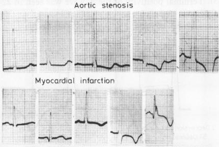
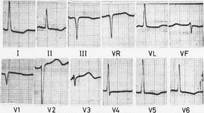
In routine reporting of electrocardiograms, a frequent problem is presented by the presence of repolarisation abnormalities (ST depression and/or T wave inversion) in the lateral leads without the accepted QRS voltage criterion of left ventricular hypertrophy. To help resolve this problem, the electrocardiograms of 41 patients with severe aortic stenosis who had no evidence of coronary disease were compared with the electrocardiograms of 20 patients with lateral myocardial infarction who had no clinical evidence of left ventricular hypertrophy. Nine of the patients with aortic stenosis were found to show repolarisation abnormalities in the lateral leads without the standard voltage criterion of left ventricular hypertrophy. The repolarisation pattern of aortic stenosis could frequently be distinguished from that of coronary disease by the presence of one or more of the following five features: depression of the J point, asymmetry of the T wave with rapid return to the baseline, terminal positivity of the T wave ("over-shoot"), T inversion in V6 greater than 3 mm, and T inversion greater in V6 than in V4.
在常规心电图报告中,常出现这样一个问题:在侧壁导联存在复极异常(ST段压低和/或T波倒置),却未达到左心室肥厚公认的QRS电压标准。为帮助解决这一问题,将41例无冠心病证据的重度主动脉瓣狭窄患者的心电图与20例无左心室肥厚临床证据的侧壁心肌梗死患者的心电图进行了比较。发现9例主动脉瓣狭窄患者在侧壁导联出现复极异常,但未达到左心室肥厚的标准电压标准。主动脉瓣狭窄的复极模式通常可通过以下五个特征中的一个或多个与冠心病的复极模式相区分:J点压低、T波不对称且快速回到基线、T波终末正向(“超射”)、V6导联T波倒置大于3mm以及V6导联T波倒置大于V4导联。