• 文献检索
  • 文档翻译
  • 深度研究
  • 学术资讯
  • Suppr Zotero 插件Zotero 插件
  • 邀请有礼
  • 套餐&价格
  • 历史记录
应用&插件
Suppr Zotero 插件Zotero 插件浏览器插件Mac 客户端Windows 客户端微信小程序
定价
高级版会员购买积分包购买API积分包
服务
文献检索文档翻译深度研究API 文档MCP 服务
关于我们
关于 Suppr公司介绍联系我们用户协议隐私条款
关注我们

Suppr 超能文献

核心技术专利:CN118964589B侵权必究
粤ICP备2023148730 号-1Suppr @ 2026

文献检索

告别复杂PubMed语法,用中文像聊天一样搜索,搜遍4000万医学文献。AI智能推荐,让科研检索更轻松。

立即免费搜索

文件翻译

保留排版,准确专业,支持PDF/Word/PPT等文件格式,支持 12+语言互译。

免费翻译文档

深度研究

AI帮你快速写综述,25分钟生成高质量综述,智能提取关键信息,辅助科研写作。

立即免费体验

左心室肥厚且冠状动脉正常患者的ST-T异常的可变模式。

Variable patterns of ST-T abnormalities in patients with left ventricular hypertrophy and normal coronary arteries.

作者信息

Huwez F U, Pringle S D, Macfarlane P W

机构信息

University Department of Medical Cardiology, Royal Infirmary, Glasgow.

出版信息

Br Heart J. 1992 Apr;67(4):304-7. doi: 10.1136/hrt.67.4.304.

DOI:10.1136/hrt.67.4.304
PMID:1389704
原文链接:https://pmc.ncbi.nlm.nih.gov/articles/PMC1024837/
Abstract

BACKGROUND

Classically, the ST-T configuration in the electrocardiogram of patients with left ventricular hypertrophy is said to have a typical pattern of ST depression together with asymmetrical T wave inversion (the so-called left ventricular strain pattern). However, many patients with left ventricular hypertrophy may also have ischaemic heart disease. To revise the electrocardiographic criteria for left ventricular hypertrophy the ST-T configuration in patients with left ventricular hypertrophy documented by echocardiography and with normal coronary arteries was assessed.

METHODS

24 patients were selected for this study. All had left ventricular hypertrophy documented by echocardiography, normal coronary arteries by cardiac catheterisation, and ST and/or T wave abnormalities in the lateral leads of their electrocardiogram. There were eight patients with aortic valve disease and 16 with hypertension who had coronary angiography as part of an investigation into the risk factors of sudden cardiac death caused by hypertensive left ventricular hypertrophy. No patient was receiving digitalis preparations or had electrolyte disturbances, and none had a previous myocardial infarction or ventricular conduction defect.

RESULTS

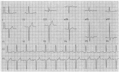
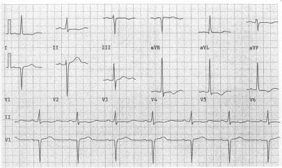
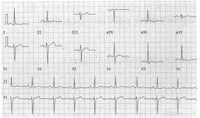
Typical electrocardiographic evidence of left ventricular strain was found in approximately two thirds (63%) of patients and 95% of this subgroup had asymmetrical T wave inversion. Flat ST segment depression, with or without T wave inversion or isolated T wave inversion (symmetrical or asymmetrical) in the anterolateral leads, was seen in the remaining 37% of patients.

CONCLUSIONS

These findings indicate that left ventricular hypertrophy without coronary artery disease can cause variable types of ST-T abnormalities in the anterolateral leads including the typical left ventricular strain pattern and non-specific ST-T changes. Non-specific abnormalities could not be distinguished from those of coronary artery disease and may adversely affect the accuracy of the electrocardiographic criteria for the diagnosis of left ventricular hypertrophy because they do not accord with the criteria for left ventricular strain.

摘要

背景

传统观点认为,左心室肥厚患者心电图的ST-T形态具有典型模式,即ST段压低伴不对称T波倒置(即所谓的左心室劳损图形)。然而,许多左心室肥厚患者也可能患有缺血性心脏病。为了修订左心室肥厚的心电图诊断标准,我们对经超声心动图证实为左心室肥厚且冠状动脉正常的患者的ST-T形态进行了评估。

方法

本研究选取了24例患者。所有患者均经超声心动图证实有左心室肥厚,经心导管检查冠状动脉正常,且心电图侧导联有ST段和/或T波异常。其中8例患有主动脉瓣疾病,16例患有高血压,他们接受冠状动脉造影作为高血压性左心室肥厚所致心源性猝死危险因素调查的一部分。所有患者均未服用洋地黄制剂,无电解质紊乱,既往无心肌梗死或心室传导缺陷。

结果

约三分之二(63%)的患者出现典型的左心室劳损心电图表现,该亚组中95%的患者有不对称T波倒置。其余37%的患者在前外侧导联可见ST段压低伴或不伴T波倒置或孤立的T波倒置(对称或不对称)。

结论

这些发现表明,无冠状动脉疾病的左心室肥厚可导致前外侧导联出现多种类型的ST-T异常,包括典型的左心室劳损图形和非特异性ST-T改变。非特异性异常无法与冠状动脉疾病所致异常相区分,可能会对左心室肥厚心电图诊断标准的准确性产生不利影响,因为它们不符合左心室劳损的标准。

https://cdn.ncbi.nlm.nih.gov/pmc/blobs/584d/1024837/efd02cedf7a2/brheartj00028-0029-b.jpg
https://cdn.ncbi.nlm.nih.gov/pmc/blobs/584d/1024837/2a00e2dba4c5/brheartj00028-0028-a.jpg
https://cdn.ncbi.nlm.nih.gov/pmc/blobs/584d/1024837/09ca64f38587/brheartj00028-0028-b.jpg
https://cdn.ncbi.nlm.nih.gov/pmc/blobs/584d/1024837/c8fe07321f7d/brheartj00028-0028-c.jpg
https://cdn.ncbi.nlm.nih.gov/pmc/blobs/584d/1024837/175e27e03fd7/brheartj00028-0029-a.jpg
https://cdn.ncbi.nlm.nih.gov/pmc/blobs/584d/1024837/efd02cedf7a2/brheartj00028-0029-b.jpg

相似文献

1
Variable patterns of ST-T abnormalities in patients with left ventricular hypertrophy and normal coronary arteries.左心室肥厚且冠状动脉正常患者的ST-T异常的可变模式。
Br Heart J. 1992 Apr;67(4):304-7. doi: 10.1136/hrt.67.4.304.
2
Prevalence and prognosis of electrocardiographic left ventricular hypertrophy, ST segment depression and negative T-wave; the Copenhagen City Heart Study.心电图左心室肥厚、ST段压低和T波倒置的患病率及预后;哥本哈根城市心脏研究
Eur Heart J. 2002 Feb;23(4):315-24. doi: 10.1053/euhj.2001.2774.
3
[Changes in ST segment on ECG of hypertensive patients].[高血压患者心电图ST段的变化]
Ter Arkh. 2008;80(5):67-73.
4
Electrocardiogram of pure left ventricular hypertrophy and its differentiation from lateral ischaemia.单纯左心室肥厚的心电图及其与侧壁心肌缺血的鉴别
Br Heart J. 1981 Sep;46(3):285-9. doi: 10.1136/hrt.46.3.285.
5
Electrocardiographic abnormalities in apparently healthy men and the risk of sudden death.貌似健康男性的心电图异常与猝死风险
Drugs. 1984 Oct;28 Suppl 1:28-45. doi: 10.2165/00003495-198400281-00004.
6
[Electrocardiographic patterns in hypertensive patients without atherosclerotic coronary disease. Influence of the left ventricular mass].[无动脉粥样硬化性冠状动脉疾病的高血压患者的心电图模式。左心室质量的影响]
Med Clin (Barc). 1995 May 20;104(19):732-6.
7
ST segment/heart rate hysteresis improves the diagnostic accuracy of ECG stress test for coronary artery disease in patients with left ventricular hypertrophy.ST段/心率滞后现象可提高心电图负荷试验对左心室肥厚患者冠心病的诊断准确性。
Eur J Prev Cardiol. 2016 Oct;23(15):1632-9. doi: 10.1177/2047487316655259. Epub 2016 Jun 27.
8
Prevalence and natural history of electrocardiographic left ventricular hypertrophy.心电图左心室肥厚的患病率及自然病史。
Am J Med. 1983 Sep 26;75(3A):4-11. doi: 10.1016/0002-9343(83)90111-0.
9
T-wave changes in patients with hemodynamic evidence of systolic or diastolic overload of the left ventricle: a retrospective study on 168 patients with isolated chronic aortic valve disease.有左心室收缩或舒张负荷过重血流动力学证据患者的T波改变:一项对168例单纯慢性主动脉瓣疾病患者的回顾性研究
Int J Cardiol. 1987 Feb;14(2):137-43. doi: 10.1016/0167-5273(87)90003-9.
10
Giant T wave inversion as a manifestation of asymmetrical apical hypertrophy (AAH) of the left ventricle. Echocardiographic and ultrasono-cardiotomographic study.巨大T波倒置作为左心室不对称性心尖肥厚(AAH)的一种表现。超声心动图和超声心动断层扫描研究。
Jpn Heart J. 1976 Sep;17(5):611-29. doi: 10.1536/ihj.17.611.

引用本文的文献

1
Improvement of electrocardiographic diagnostic accuracy of left ventricular hypertrophy using a Machine Learning approach.采用机器学习方法提高左心室肥厚的心电图诊断准确性。
PLoS One. 2020 May 13;15(5):e0232657. doi: 10.1371/journal.pone.0232657. eCollection 2020.
2
Association of isolated minor nonspecific ST-T abnormalities with left ventricular hypertrophy and diastolic dysfunction.孤立性小非特异性 ST-T 异常与左心室肥厚和舒张功能障碍的关系。
Sci Rep. 2018 Jun 8;8(1):8791. doi: 10.1038/s41598-018-27028-6.
3
How Dangerous Is Hyperkalemia?

本文引用的文献

1
ELECTROCARDIOGRAPHIC MANIFESTATIONS OF VENTRICULAR HYPERTROPHY; A COMPUTER STUDY OF ECG-ANATOMIC CORRELATIONS IN 319 CASES.心室肥厚的心电图表现;319例心电图与解剖学相关性的计算机研究
Am Heart J. 1964 Aug;68:173-82. doi: 10.1016/0002-8703(64)90038-9.
2
Repolarization abnormalities of left ventricular hypertrophy. Clinical, echocardiographic and hemodynamic correlates.左心室肥厚的复极异常。临床、超声心动图及血流动力学相关性
J Electrocardiol. 1982 Jan;15(1):47-53. doi: 10.1016/s0022-0736(82)80044-7.
3
Electrocardiogram of pure left ventricular hypertrophy and its differentiation from lateral ischaemia.
高钾血症有多危险?
J Am Soc Nephrol. 2017 Nov;28(11):3155-3165. doi: 10.1681/ASN.2016121344. Epub 2017 Aug 4.
4
Prevalence of electrocardiographic abnormalities based on hypertension severity and blood pressure levels: the Reasons for Geographic and Racial Differences in Stroke study.基于高血压严重程度和血压水平的心电图异常患病率:中风地理和种族差异原因研究
J Am Soc Hypertens. 2016 Sep;10(9):702-713.e4. doi: 10.1016/j.jash.2016.06.033. Epub 2016 Jun 27.
5
Hemodynamic responses and upper airway morbidity following tracheal intubation in patients with hypertension: conventional laryngoscopy versus an intubating laryngeal mask airway.高血压患者气管插管后的血流动力学反应和上呼吸道并发症:常规喉镜与插管型喉罩气道。
Clinics (Sao Paulo). 2012;67(1):49-54. doi: 10.6061/clinics/2012(01)08.
6
Electrocardiographic left ventricular hypertrophy with strain pattern: prevalence, mechanisms and prognostic implications.伴有应变模式的心电图左心室肥厚:患病率、机制及预后意义。
Cardiovasc J Afr. 2008 Jan-Feb;19(1):39-45.
7
Prognosis following acute myocardial infarction in patients with ST-T abnormalities on electrocardiograms obtained before myocardial infarction.心肌梗死前心电图有ST-T异常的患者急性心肌梗死后的预后
Clin Cardiol. 2001 Feb;24(2):107-13. doi: 10.1002/clc.4960240203.
8
Electrocardiographic ST segment elevation in adults with chest pain.成人胸痛患者的心电图ST段抬高
J Accid Emerg Med. 1999 Nov;16(6):433-9. doi: 10.1136/emj.16.6.433.
9
Detection of electrocardiographic 'left ventricular strain' using neural nets.
Med Biol Eng Comput. 1993 Jul;31(4):343-8. doi: 10.1007/BF02446686.
单纯左心室肥厚的心电图及其与侧壁心肌缺血的鉴别
Br Heart J. 1981 Sep;46(3):285-9. doi: 10.1136/hrt.46.3.285.
4
Standardization of M-mode echocardiographic left ventricular anatomic measurements.M型超声心动图左心室解剖测量的标准化
J Am Coll Cardiol. 1984 Dec;4(6):1222-30. doi: 10.1016/s0735-1097(84)80141-2.
5
Electrocardiographic left ventricular hypertrophy and risk of coronary heart disease. The Framingham study.心电图左心室肥厚与冠心病风险。弗明汉姆研究。
Ann Intern Med. 1970 Jun;72(6):813-22. doi: 10.7326/0003-4819-72-6-813.
6
Pathophysiologic assessment of left ventricular hypertrophy and strain in asymptomatic patients with essential hypertension.
J Am Coll Cardiol. 1989 May;13(6):1377-81. doi: 10.1016/0735-1097(89)90314-8.
7
Echocardiographic determination of left ventricular mass in man. Anatomic validation of the method.
Circulation. 1977 Apr;55(4):613-8. doi: 10.1161/01.cir.55.4.613.