• 文献检索
  • 文档翻译
  • 深度研究
  • 学术资讯
  • Suppr Zotero 插件Zotero 插件
  • 邀请有礼
  • 套餐&价格
  • 历史记录
应用&插件
Suppr Zotero 插件Zotero 插件浏览器插件Mac 客户端Windows 客户端微信小程序
定价
高级版会员购买积分包购买API积分包
服务
文献检索文档翻译深度研究API 文档MCP 服务
关于我们
关于 Suppr公司介绍联系我们用户协议隐私条款
关注我们

Suppr 超能文献

核心技术专利:CN118964589B侵权必究
粤ICP备2023148730 号-1Suppr @ 2026

文献检索

告别复杂PubMed语法,用中文像聊天一样搜索,搜遍4000万医学文献。AI智能推荐,让科研检索更轻松。

立即免费搜索

文件翻译

保留排版,准确专业,支持PDF/Word/PPT等文件格式,支持 12+语言互译。

免费翻译文档

深度研究

AI帮你快速写综述,25分钟生成高质量综述,智能提取关键信息,辅助科研写作。

立即免费体验

The postnatal development of blood vessels in the optic nerve of normotensive and hypertensive rats.

作者信息

Scott T M, Foote J

出版信息

J Anat. 1984 Jun;138 ( Pt 4)(Pt 4):635-42.

PMID:6746402
原文链接:https://pmc.ncbi.nlm.nih.gov/articles/PMC1164349/
Abstract

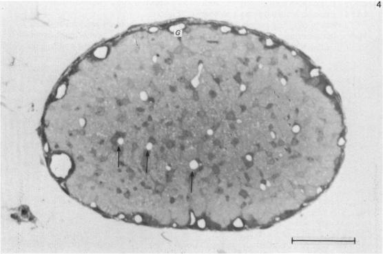
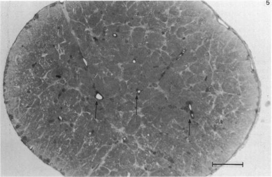
The blood vessels in the optic nerve of normotensive and hypertensive rats have been examined at 2, 4, 8 and 12 weeks of age. The pattern of development was found to be different in the two strains, with the number of blood vessels in the hypertensive rat optic nerve being lower at 2 weeks, but greater at 12 weeks than the normotensive rat. There appeared to be no correlation between vascularity and either myelination or changes in the fibre diameter spectrum at the ages studied. It is concluded that while the cause of the increased vascularity of the optic nerve in hypertensive rats is not known, it appears to be without effect in the structural development of the optic nerve.

摘要
https://cdn.ncbi.nlm.nih.gov/pmc/blobs/bd52/1164349/985c7d5d3699/janat00204-0038-d.jpg
https://cdn.ncbi.nlm.nih.gov/pmc/blobs/bd52/1164349/5dc9eed609c6/janat00204-0036-a.jpg
https://cdn.ncbi.nlm.nih.gov/pmc/blobs/bd52/1164349/616a8458a354/janat00204-0036-b.jpg
https://cdn.ncbi.nlm.nih.gov/pmc/blobs/bd52/1164349/5308ba647bea/janat00204-0038-a.jpg
https://cdn.ncbi.nlm.nih.gov/pmc/blobs/bd52/1164349/0bc29a53d09e/janat00204-0038-b.jpg
https://cdn.ncbi.nlm.nih.gov/pmc/blobs/bd52/1164349/93c8faba176a/janat00204-0038-c.jpg
https://cdn.ncbi.nlm.nih.gov/pmc/blobs/bd52/1164349/985c7d5d3699/janat00204-0038-d.jpg

相似文献

1
The postnatal development of blood vessels in the optic nerve of normotensive and hypertensive rats.
J Anat. 1984 Jun;138 ( Pt 4)(Pt 4):635-42.
2
A quantitative morphological study of interstrain variation in the developing rat optic nerve.发育中大鼠视神经株间变异的定量形态学研究
J Comp Neurol. 1986 Mar 8;245(2):160-6. doi: 10.1002/cne.902450203.
3
Quantitative study of the development of the optic nerve in rats reared in the dark during early postnatal life.对出生后早期生活在黑暗环境中的大鼠视神经发育的定量研究。
J Anat. 1991 Feb;174:37-47.
4
[Stages in the postnatal maturation of the optic nerve: morphometric changes during the critical period].[视神经产后成熟的阶段:关键期的形态计量学变化]
Arch Neurobiol (Madr). 1989 Jan-Feb;52(1):1-8.
5
Postnatal development of the optic nerve in (C57BL x CBA)F1 hybrid mice: general changes in morphometric parameters.(C57BL×CBA)F1杂交小鼠视神经的出生后发育:形态学参数的一般变化
J Anat. 1996 Aug;189 ( Pt 1)(Pt 1):117-25.
6
Developmental changes in the fibre population of the optic nerve follow an avian/mammalian-like pattern in the turtle Mauremys leprosa.
Brain Res. 2006 Oct 3;1113(1):74-85. doi: 10.1016/j.brainres.2006.07.040. Epub 2006 Aug 28.
7
Postnatal development of microtubules and neurofilaments in the rat optic nerve: a quantitative study.大鼠视神经中微管和神经丝的产后发育:一项定量研究。
J Comp Neurol. 1987 Sep 22;263(4):613-7. doi: 10.1002/cne.902630412.
8
The effect of neonatal monocular enucleation on the optic nerves of the rat.新生大鼠单眼摘除对视神经的影响。
J Anat. 1991 Feb;174:27-35.
9
Vascularization of the human embryonic optic nerve.人类胚胎视神经的血管形成
J Hirnforsch. 1987;28(6):615-24.
10
Stereological analysis of the tunica media of the aorta and renal artery during the development of hypertension in the spontaneously hypertensive rat.自发性高血压大鼠高血压发展过程中主动脉和肾动脉中膜的体视学分析
J Anat. 1981 Dec;133(Pt 4):513-26.

引用本文的文献

1
Novel morphological features of developing white matter pericytes and rapid scavenging of reactive oxygen species in the neighbouring endothelia.发育中的脑白质周细胞的新形态特征及其邻近内皮细胞中活性氧的快速清除。
J Anat. 2011 Jul;219(1):65-77. doi: 10.1111/j.1469-7580.2011.01373.x. Epub 2011 Apr 12.
2
Vascular and neural changes in the rat optic nerve following induction of diabetes with streptozotocin.
J Anat. 1986 Feb;144:145-52.

本文引用的文献

1
Pattern of myelination and distribution of neuroglial cells along the developing optic system of the rat and rabbit.大鼠和家兔发育中的视觉系统中神经髓鞘形成模式及神经胶质细胞分布
J Comp Neurol. 1980 May 15;191(2):237-53. doi: 10.1002/cne.901910207.
2
Stereological analysis of the tunica media of the aorta and renal artery during the development of hypertension in the spontaneously hypertensive rat.自发性高血压大鼠高血压发展过程中主动脉和肾动脉中膜的体视学分析
J Anat. 1981 Dec;133(Pt 4):513-26.
3
A quantitative study of vascularisation of the prenatal rabbit spinal cord.
J Anat. 1982 Aug;135(Pt 1):89-96.
4
Rat optic nerve: electrophysiological, pharmacological and anatomical studies during development.大鼠视神经:发育过程中的电生理、药理学及解剖学研究
Brain Res. 1982 Mar;255(3):371-86. doi: 10.1016/0165-3806(82)90005-0.
5
Comparison of microvascular pressures in normal and spontaneously hypertensive rats.
Microvasc Res. 1977 Jan;13(1):125-30. doi: 10.1016/0026-2862(77)90121-2.