Rognum T, Elgjo K, Brandtzaeg P, Orjasaeter H, Bergan A
J Clin Pathol. 1982 Sep;35(9):922-33. doi: 10.1136/jcp.35.9.922.
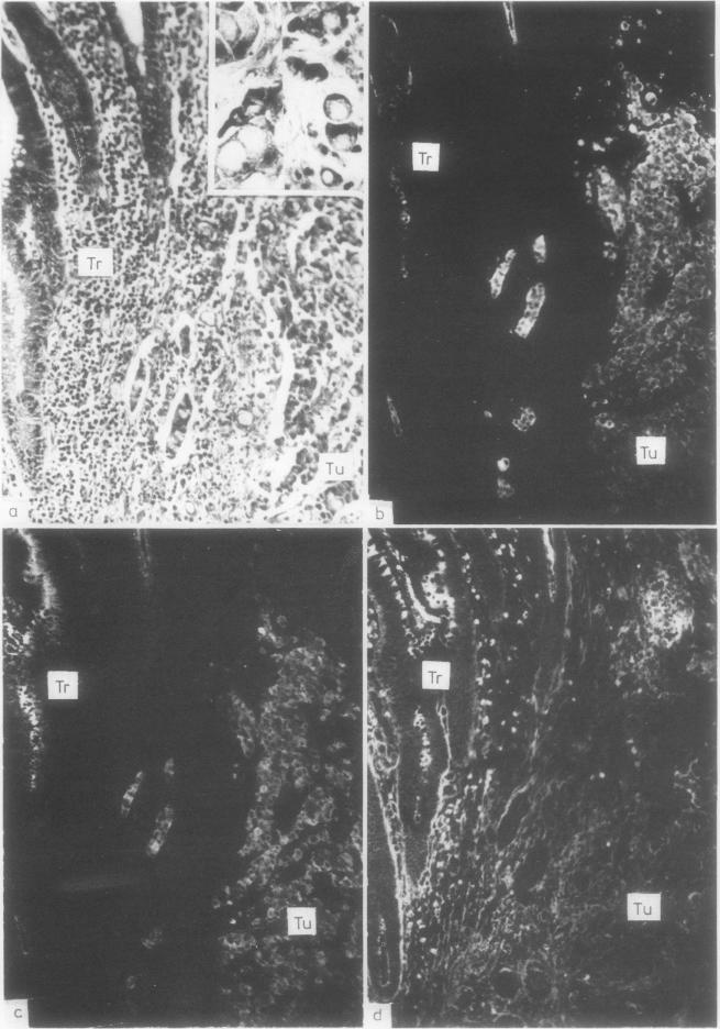
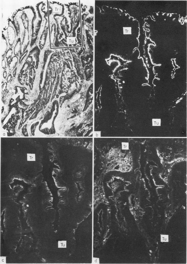
Carcinoembryonic antigen (CEA), secretory component (SC), and epithelial IgA were traced by paired immunofluorescence staining in 102 large bowel carcinomas from 99 patients. The immunohistochemical results were evaluated semiquantitatively in relation to histological tumour grade, clinicopathological stage, and preoperative plasma CEA concentration. CEA expression was significantly increased (p less than 0.05) in the following order: histologically normal colon mucosa, transitional mucosa adjacent to tumours, neoplastic epithelium; the reverse was true for the expression of SC and epithelial IgA (p less than 0.01). CEA was significantly more abundant in the moderately and poorly differentiated tumors than in the well differentiated ones (p less than 0.05), whereas the latter showed better expression of SC (p less than 0.05) and epithelial IgA (p approximately 0.06). In the transitional mucosa, CEA staining tended to be inversely related to histological tumour grade, whereas SC and epithelial IgA were significantly better seen in this zone when the adjacent tumour was well differentiated than when it was moderately or poorly differentiated (p less than 0.01). Furthermore, the expression of SC and epithelial IgA in the transitional mucosa decreased with increasing invasiveness of the tumours, whereas the opposite relation was indicated for CEA expression. Plasma CEA concentrations were not clearly correlated with histological levels than the localised well differentiated tumours tended to be associated with lower levels than the localised moderately differentiated ones (p approximately 0.06). Moreover, the latter variety was associated with lower plasma CEA concentrations than disseminated tumours of comparable differentiation (p less than 0.01).
采用配对免疫荧光染色法对99例患者的102例大肠癌组织中的癌胚抗原(CEA)、分泌成分(SC)和上皮型IgA进行检测。根据组织学肿瘤分级、临床病理分期和术前血浆CEA浓度对免疫组化结果进行半定量评估。CEA表达按以下顺序显著增加(p<0.05):组织学正常的结肠黏膜、肿瘤旁过渡黏膜、肿瘤上皮;而SC和上皮型IgA的表达情况则相反(p<0.01)。中分化和低分化肿瘤中的CEA明显比高分化肿瘤丰富(p<0.05),而高分化肿瘤中SC(p<0.05)和上皮型IgA(p约为0.06)的表达更好。在过渡黏膜中,CEA染色与组织学肿瘤分级呈负相关,而当相邻肿瘤为高分化时,该区域的SC和上皮型IgA比中分化或低分化时明显更易观察到(p<0.01)。此外,过渡黏膜中SC和上皮型IgA的表达随肿瘤侵袭性增加而降低,而CEA表达则呈相反关系。血浆CEA浓度与组织学分级无明显相关性,局限性高分化肿瘤的血浆CEA浓度往往低于局限性中分化肿瘤(p约为0.06)。此外,与分化程度相当的播散性肿瘤相比,后者的血浆CEA浓度更低(p<0.01)。