Fehlmann M, Carpentier J L, Van Obberghen E, Freychet P, Thamm P, Saunders D, Brandenburg D, Orci L
Proc Natl Acad Sci U S A. 1982 Oct;79(19):5921-5. doi: 10.1073/pnas.79.19.5921.
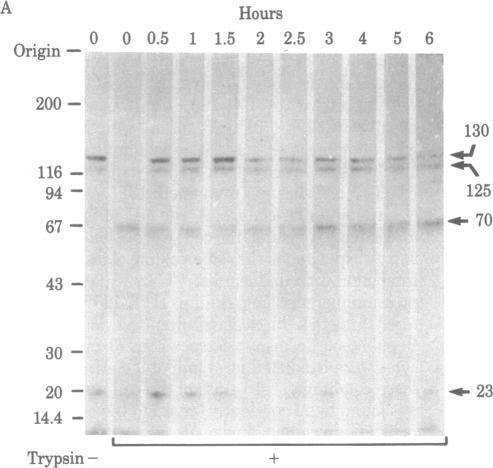
We have followed the fate of cell surface insulin receptors in isolated rat hepatocytes by both a biochemical and a morphological approach. Hepatocytes were labeled with the photoreactive and biologically active 125I-labeled insulin analogue, [2-nitro-4-azidophenylacetylB2]des-PheB1-insulin, under conditions that allow for minimal internalization (2 hr at 15 degrees C). Analysis of the cell-associated radioactivity by NaDodSO4/polyacrylamide gel electrophoresis under reducing conditions followed by autoradiography revealed the specific labeling of a major insulin receptor subunit with Mr 130,000 and a minor degradation product with Mr 125,000. When the cells were exposed at 15 degrees C to trypsin at the end of the association period, these two bands were no longer observed, indicating that the labeled receptors were at the cell surface. This trypsin sensitivity of the receptor disappeared within 30-60 min of incubation of the cells at 37 degrees C, reflecting the internalization of the hormone-receptor complexes. Over the subsequent 4 hr of incubation, this was followed by a progressive reappearance of the receptor complexes at the cell surface, as indicated by the recovery of trypsin sensitivity of the labeled insulin receptors. An identical (both chronologically and quantitatively) journey of the insulin receptors was observed when the labeled material was studied by quantitative electron microscopic autoradiography. Thus, when the cells were incubated at 37 degrees C there was a rapid decrease (30-60 min) in the percentage of autoradiographic grains associated with the plasma membrane, followed by a progressive increase in this percentage over the subsequent 4 hr of incubation. In conclusion, using a biochemical and morphological approach to trace the photoaffinity-labeled insulin receptor, we have shown that the internalized hormone-receptor complex is recycled back to the cell surface.
我们通过生化和形态学方法追踪了分离的大鼠肝细胞中细胞表面胰岛素受体的命运。在允许最小内化的条件下(15℃孵育2小时),用光反应性和生物活性的125I标记胰岛素类似物[2-硝基-4-叠氮基苯乙酰B2]去-PheB1-胰岛素标记肝细胞。在还原条件下通过NaDodSO4/聚丙烯酰胺凝胶电泳分析细胞相关放射性,随后进行放射自显影,结果显示主要胰岛素受体亚基Mr 130,000和次要降解产物Mr 125,000有特异性标记。当在结合期结束时将细胞在15℃暴露于胰蛋白酶时,这两条带不再出现,表明标记的受体位于细胞表面。受体的这种胰蛋白酶敏感性在细胞于37℃孵育30 - 60分钟内消失,这反映了激素-受体复合物的内化。在随后的4小时孵育过程中,标记的胰岛素受体对胰蛋白酶敏感性的恢复表明,受体复合物在细胞表面逐渐重新出现。当通过定量电子显微镜放射自显影研究标记物质时,观察到胰岛素受体经历了相同的(时间和数量上)过程。因此,当细胞在37℃孵育时,与质膜相关的放射自显影颗粒百分比迅速下降(30 - 60分钟),随后在接下来的4小时孵育过程中该百分比逐渐增加。总之,使用生化和形态学方法追踪光亲和标记的胰岛素受体,我们已经表明内化的激素-受体复合物会再循环回到细胞表面。