• 文献检索
  • 文档翻译
  • 深度研究
  • 学术资讯
  • Suppr Zotero 插件Zotero 插件
  • 邀请有礼
  • 套餐&价格
  • 历史记录
应用&插件
Suppr Zotero 插件Zotero 插件浏览器插件Mac 客户端Windows 客户端微信小程序
定价
高级版会员购买积分包购买API积分包
服务
文献检索文档翻译深度研究API 文档MCP 服务
关于我们
关于 Suppr公司介绍联系我们用户协议隐私条款
关注我们

Suppr 超能文献

核心技术专利:CN118964589B侵权必究
粤ICP备2023148730 号-1Suppr @ 2026

文献检索

告别复杂PubMed语法,用中文像聊天一样搜索,搜遍4000万医学文献。AI智能推荐,让科研检索更轻松。

立即免费搜索

文件翻译

保留排版,准确专业,支持PDF/Word/PPT等文件格式,支持 12+语言互译。

免费翻译文档

深度研究

AI帮你快速写综述,25分钟生成高质量综述,智能提取关键信息,辅助科研写作。

立即免费体验

鲁氏毛霉节孢子的形成及超微结构

Formation and ultrastructure of Mucor rouxii arthrospores.

作者信息

Barrera C R

出版信息

J Bacteriol. 1983 Aug;155(2):886-95. doi: 10.1128/jb.155.2.886-895.1983.

DOI:10.1128/jb.155.2.886-895.1983
PMID:6874646
原文链接:https://pmc.ncbi.nlm.nih.gov/articles/PMC217764/
Abstract

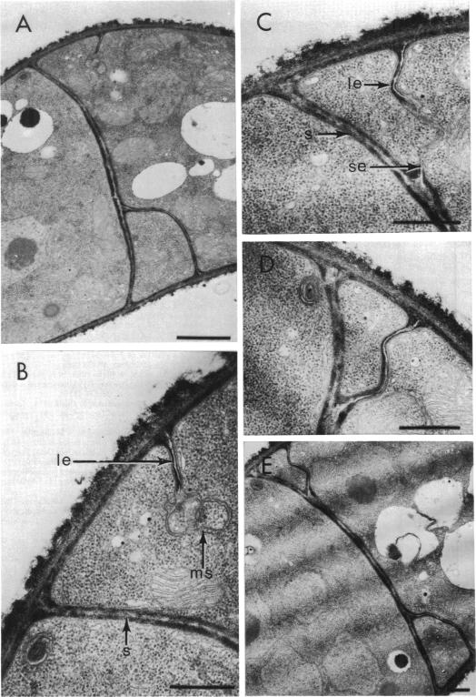
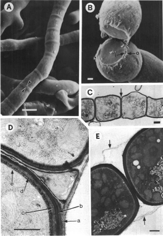
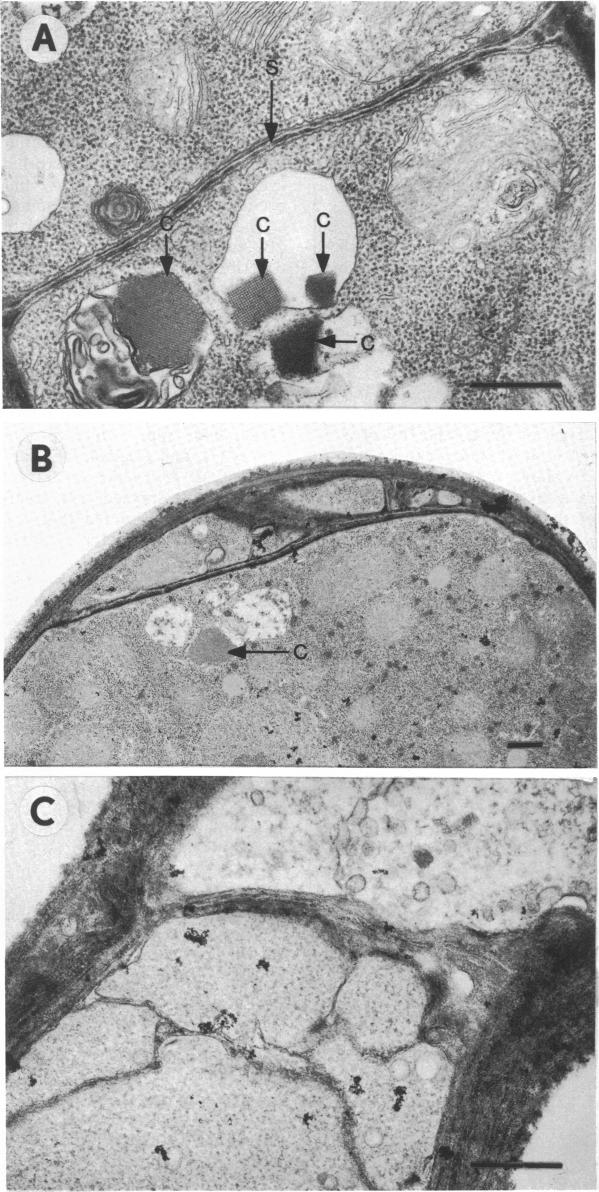
The formation of arthrospores in Mucor rouxii was studied by transmission and scanning electron microscopy and light microscopy. The arthrospores formed in a random manner in terminal and internal regions of the hyphae. The earliest appearance of the arthrospores was seen by scanning electron microscopy as compartments delineated by double ridges. These ridges probably corresponded to the site of septal wall formation. The elongated compartments varied considerably in size. As the arthrospores matured, they tended to separate as a result of a gradual change in the shape of the arthrospores to a nearly spherical form and also as the result of simultaneous degradation of the outermost cell wall layer. The mature arthrospores were surrounded by a complex cell wall consisting of at least three distinct layers in addition to the original hyphal cell wall. Crystal-like structures were seen in the cytoplasm of some of the arthrospores in addition to the usual organelles such as mitochondria, nuclei, and ribosomes. Septum formation by centripetal cell wall growth from the lateral hyphal wall was documented by transmission electron microscopy. However, evidence was also found which suggested that not all internal cell wall development in the fungal hyphae during arthrosporogenesis necessarily led to the formation of mature arthrospores.

摘要

通过透射电子显微镜、扫描电子显微镜和光学显微镜对鲁氏毛霉中节孢子的形成进行了研究。节孢子在菌丝的末端和内部区域随机形成。扫描电子显微镜观察到节孢子最早的形态是由双嵴界定的间隔区。这些嵴可能对应于隔壁形成的部位。拉长的间隔区大小差异很大。随着节孢子的成熟,由于节孢子形状逐渐变为近球形,以及最外层细胞壁层同时降解,它们倾向于分离。成熟的节孢子除了原来的菌丝细胞壁外,还被一层由至少三层不同层组成的复杂细胞壁所包围。除了线粒体、细胞核和核糖体等常见细胞器外,在一些节孢子的细胞质中还可见到晶体状结构。透射电子显微镜记录了从菌丝侧壁向心生长形成隔膜的过程。然而,也发现有证据表明,在节孢子形成过程中,真菌菌丝内并非所有的内部细胞壁发育都必然导致成熟节孢子的形成。

https://cdn.ncbi.nlm.nih.gov/pmc/blobs/8558/217764/519b1962f629/jbacter00243-0457-a.jpg
https://cdn.ncbi.nlm.nih.gov/pmc/blobs/8558/217764/2dbaaada4396/jbacter00243-0452-a.jpg
https://cdn.ncbi.nlm.nih.gov/pmc/blobs/8558/217764/edcf52f7b5d9/jbacter00243-0453-a.jpg
https://cdn.ncbi.nlm.nih.gov/pmc/blobs/8558/217764/bea61376425c/jbacter00243-0454-a.jpg
https://cdn.ncbi.nlm.nih.gov/pmc/blobs/8558/217764/67abd8a1c993/jbacter00243-0456-a.jpg
https://cdn.ncbi.nlm.nih.gov/pmc/blobs/8558/217764/519b1962f629/jbacter00243-0457-a.jpg

相似文献

1
Formation and ultrastructure of Mucor rouxii arthrospores.鲁氏毛霉节孢子的形成及超微结构
J Bacteriol. 1983 Aug;155(2):886-95. doi: 10.1128/jb.155.2.886-895.1983.
2
Development of arthrospores of Trichophyton mentagrophytes.须癣毛癣菌关节孢子的发育
Infect Immun. 1977 Mar;15(3):958-71. doi: 10.1128/iai.15.3.958-971.1977.
3
Differentition of mutants of Cephalosporium acremonium in complex medium: the formation of unicellular arthrospores and their germination.顶头孢霉突变体在复合培养基中的分化:单细胞节孢子的形成及其萌发
Can J Microbiol. 1975 Dec;21(12):1981-96. doi: 10.1139/m75-286.
4
Ultrastructure of spore formation in Paracoccidioides brasiliensis.巴西副球孢子菌孢子形成的超微结构
J Med Vet Mycol. 1986 Jun;24(3):203-10.
5
Spore morphology and ultrastructure of an Ascosphaera apis strain from the honeybees (Apis mellifera) in southwest China.来自中国西南部蜜蜂(西方蜜蜂)的蜜蜂球囊菌菌株的孢子形态和超微结构
Mycologia. 2018 Mar-Apr;110(2):325-338. doi: 10.1080/00275514.2018.1442084. Epub 2018 May 31.
6
[Composition of sterols from arthrospores and mycelia of the fungus Mucor hiemalis].[来自冬生毛霉真菌的节孢子和菌丝体中甾醇的组成]
Mikrobiologiia. 2002 Jul-Aug;71(4):475-81.
7
Chitosomes and chitin synthetase in the asexual life cycle of Mucor rouxii: spores, mycelium and yeast cells.鲁氏毛霉无性生命周期中的几丁质体和几丁质合成酶:孢子、菌丝体和酵母细胞。
J Gen Microbiol. 1991 Jun;137(6):1241-52. doi: 10.1099/00221287-137-6-1241.
8
Characterization of the Mucor circinelloides life cycle by on-line image analysis.通过在线图像分析对卷枝毛霉生命周期的表征。
J Appl Microbiol. 2003;95(5):1152-60. doi: 10.1046/j.1365-2672.2003.02098.x.
9
Induction of yeast-like development in Mucor by carbon dioxide.二氧化碳诱导毛霉产生酵母样发育。
J Bacteriol. 1962 Oct;84(4):829-40. doi: 10.1128/jb.84.4.829-840.1962.
10
Ultrastructure of spore development in Scutellospora heterogama.异形盾巨孢囊霉孢子发育的超微结构
Mycorrhiza. 2007 Jul;17(5):395-403. doi: 10.1007/s00572-007-0112-y. Epub 2007 Feb 20.

引用本文的文献

1
a novel dimorphic species resembling in a clinical sample: questions on ecological strategy.一株在临床样本中类似 的新型二相性物种:关于生态策略的问题。
mBio. 2024 Aug 14;15(8):e0014424. doi: 10.1128/mbio.00144-24. Epub 2024 Jul 2.
2
Models of cell differentiation in conidial fungi.分生孢子真菌中的细胞分化模型。
Microbiol Rev. 1986 Jun;50(2):95-132. doi: 10.1128/mr.50.2.95-132.1986.
3
Mucor dimorphism.毛霉的二态性。

本文引用的文献

1
Nutrition, growth, and morphogenesis of Mucor rouxii.鲁氏毛霉的营养、生长与形态发生
J Bacteriol. 1962 Oct;84(4):841-58. doi: 10.1128/jb.84.4.841-858.1962.
2
Induction of yeast-like development in Mucor by carbon dioxide.二氧化碳诱导毛霉产生酵母样发育。
J Bacteriol. 1962 Oct;84(4):829-40. doi: 10.1128/jb.84.4.829-840.1962.
3
Isolation, composition, and structure of cell walls of filamentous and yeast-like forms of Mucor rouxii.鲁氏毛霉丝状和酵母样形态细胞壁的分离、组成及结构
Microbiol Rev. 1991 Jun;55(2):234-58. doi: 10.1128/mr.55.2.234-258.1991.
Biochim Biophys Acta. 1962 Mar 26;58:102-19. doi: 10.1016/0006-3002(62)90822-3.
4
Role of membranes of mycelial Mucor rouxii in synthesis and secretion of cell wall matrix polymers.鲁氏毛霉菌丝体膜在细胞壁基质聚合物合成与分泌中的作用
J Bacteriol. 1981 Jan;145(1):272-9. doi: 10.1128/jb.145.1.272-279.1981.
5
[Alcohol dehydrogenase (ADH) in yeasts. III. Structural investigations on cellular ADH-crystals in Saccharomyces carlsbergensis by means of electronmicroscopy and small angle x-ray scattering (author's transl)].[酵母中的乙醇脱氢酶(ADH)。III. 通过电子显微镜和小角X射线散射对卡尔斯伯酵母细胞ADH晶体的结构研究(作者译)]
Mikroskopie. 1980 Jun;36(3-4):81-92.
6
Dimorphism in pathogenic fungi.
Crit Rev Microbiol. 1980;8(1):49-97. doi: 10.3109/10408418009085078.
7
A low-viscosity epoxy resin embedding medium for electron microscopy.一种用于电子显微镜的低粘度环氧树脂包埋介质。
J Ultrastruct Res. 1969 Jan;26(1):31-43. doi: 10.1016/s0022-5320(69)90033-1.
8
Ligand-mediated osmium binding: its application in coating biological specimens for scanning electron microscopy.配体介导的锇结合:其在扫描电子显微镜生物样本包被中的应用。
J Ultrastruct Res. 1973 Nov;45(3):254-8. doi: 10.1016/s0022-5320(73)80051-6.
9
Morphogenesis and ultrastructure of Geotrichum candidum septa.白地霉隔膜的形态发生与超微结构
J Bacteriol. 1973 Oct;116(1):447-55. doi: 10.1128/jb.116.1.447-455.1973.
10
Comparison of Coccidioides immitis arthrospore, mycelium, and spherule cell walls, and influence of growth medium on mycelial cell wall composition.粗球孢子菌关节孢子、菌丝体和球形体细胞壁的比较,以及生长培养基对菌丝体细胞壁组成的影响。
Infect Immun. 1977 Jul;17(1):91-7. doi: 10.1128/iai.17.1.91-97.1977.