• 文献检索
  • 文档翻译
  • 深度研究
  • 学术资讯
  • Suppr Zotero 插件Zotero 插件
  • 邀请有礼
  • 套餐&价格
  • 历史记录
应用&插件
Suppr Zotero 插件Zotero 插件浏览器插件Mac 客户端Windows 客户端微信小程序
定价
高级版会员购买积分包购买API积分包
服务
文献检索文档翻译深度研究API 文档MCP 服务
关于我们
关于 Suppr公司介绍联系我们用户协议隐私条款
关注我们

Suppr 超能文献

核心技术专利:CN118964589B侵权必究
粤ICP备2023148730 号-1Suppr @ 2026

文献检索

告别复杂PubMed语法,用中文像聊天一样搜索,搜遍4000万医学文献。AI智能推荐,让科研检索更轻松。

立即免费搜索

文件翻译

保留排版,准确专业,支持PDF/Word/PPT等文件格式,支持 12+语言互译。

免费翻译文档

深度研究

AI帮你快速写综述,25分钟生成高质量综述,智能提取关键信息,辅助科研写作。

立即免费体验

肿瘤组织细胞-单核细胞的超微结构鉴定:一种新开发的细胞化学技术的应用

Ultrastructural identification of neoplastic histiocytes--monocytes: an application of a newly developed cytochemical technique.

作者信息

Kim H, Pangalis G A, Payne B C, Kadin M E, Rappaport H

出版信息

Am J Pathol. 1982 Feb;106(2):204-23.

PMID:6950665
原文链接:https://pmc.ncbi.nlm.nih.gov/articles/PMC1916177/
Abstract

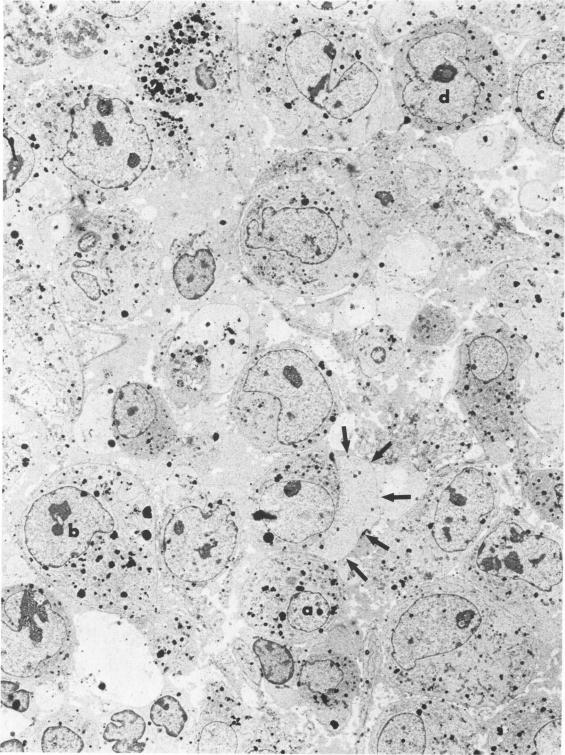
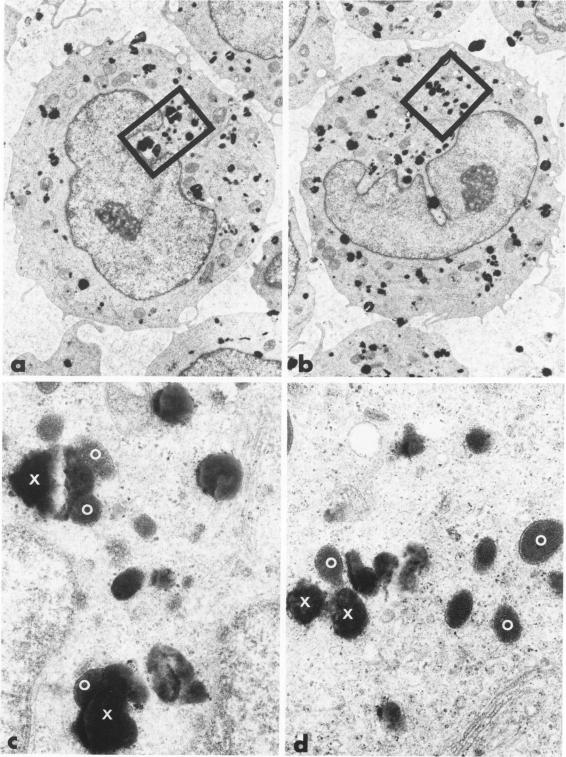
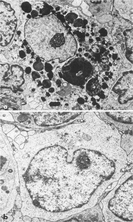
We applied a newly developed ultrastructural cytochemical technique utilizing 2-naphthyl thiol acetate (NTA) as a substrate in 3 cases of true histiocytic-monocytic malignancy and 3 controls. The enzyme NTA esterase was identified in both neoplastic and benign histiocytes-monocytes in the form of electron-dense material within the cytoplasm. The NTA esterase varied in size and shape and showed no clear relationship with cytoplasmic organelles. The distribution and pattern of staining were very similar to those of the light-microscopically demonstrated alpha-naphthyl acetate esterase reaction. The main advantages of this method are: 1) histiocytes-monocytes can be identified in the absence of lysosomes or phagocytosis; 2) unequivocal, simultaneous documentation of both histiocytic esterase and subcellular structure is possible; and 3) both blood and solid tissue specimens can be utilized. Our findings demonstrate the usefulness and applicability of ultrastructurally demonstrated NTA esterase in the study of histiocytic-monocytic malignancies.

摘要

我们应用一种新开发的超微结构细胞化学技术,以2-萘硫醇乙酸酯(NTA)为底物,对3例真性组织细胞-单核细胞恶性肿瘤患者和3例对照进行了研究。在肿瘤性和良性组织细胞-单核细胞中均鉴定出NTA酯酶,其表现为细胞质内电子致密物质的形式。NTA酯酶在大小和形状上各不相同,且与细胞质细胞器无明显关系。染色的分布和模式与光镜下显示的α-萘乙酸酯酶反应非常相似。该方法的主要优点是:1)在不存在溶酶体或吞噬作用的情况下可以识别组织细胞-单核细胞;2)可以明确、同时记录组织细胞酯酶和亚细胞结构;3)血液和实体组织标本均可使用。我们的研究结果证明了超微结构显示的NTA酯酶在组织细胞-单核细胞恶性肿瘤研究中的实用性和适用性。

https://cdn.ncbi.nlm.nih.gov/pmc/blobs/c1c8/1916177/c91bad6433fb/amjpathol00209-0085-a.jpg
https://cdn.ncbi.nlm.nih.gov/pmc/blobs/c1c8/1916177/235276348e0d/amjpathol00209-0071-a.jpg
https://cdn.ncbi.nlm.nih.gov/pmc/blobs/c1c8/1916177/377797152c67/amjpathol00209-0072-a.jpg
https://cdn.ncbi.nlm.nih.gov/pmc/blobs/c1c8/1916177/8986534478e2/amjpathol00209-0073-a.jpg
https://cdn.ncbi.nlm.nih.gov/pmc/blobs/c1c8/1916177/f795a08142a6/amjpathol00209-0074-a.jpg
https://cdn.ncbi.nlm.nih.gov/pmc/blobs/c1c8/1916177/5c978fddf32a/amjpathol00209-0076-a.jpg
https://cdn.ncbi.nlm.nih.gov/pmc/blobs/c1c8/1916177/4e8311dad354/amjpathol00209-0077-a.jpg
https://cdn.ncbi.nlm.nih.gov/pmc/blobs/c1c8/1916177/d9ea1e890abf/amjpathol00209-0078-a.jpg
https://cdn.ncbi.nlm.nih.gov/pmc/blobs/c1c8/1916177/85b05d2d8712/amjpathol00209-0079-a.jpg
https://cdn.ncbi.nlm.nih.gov/pmc/blobs/c1c8/1916177/66fbb2fdffac/amjpathol00209-0080-a.jpg
https://cdn.ncbi.nlm.nih.gov/pmc/blobs/c1c8/1916177/328f52f35f26/amjpathol00209-0081-a.jpg
https://cdn.ncbi.nlm.nih.gov/pmc/blobs/c1c8/1916177/40d916e56d16/amjpathol00209-0082-a.jpg
https://cdn.ncbi.nlm.nih.gov/pmc/blobs/c1c8/1916177/446a8346bc21/amjpathol00209-0083-a.jpg
https://cdn.ncbi.nlm.nih.gov/pmc/blobs/c1c8/1916177/c91bad6433fb/amjpathol00209-0085-a.jpg

相似文献

1
Ultrastructural identification of neoplastic histiocytes--monocytes: an application of a newly developed cytochemical technique.肿瘤组织细胞-单核细胞的超微结构鉴定:一种新开发的细胞化学技术的应用
Am J Pathol. 1982 Feb;106(2):204-23.
2
A method for the ultrastructural demonstration of non-specific esterase in human blood and lymphoid tissue.一种用于人体血液和淋巴组织中非特异性酯酶超微结构显示的方法。
Histochem J. 1980 Jan;12(1):71-86. doi: 10.1007/BF01066538.
3
Malignant histiocytosis: A light- and electron-microscopic and histochemical study.恶性组织细胞增多症:光镜、电镜及组织化学研究
Am J Surg Pathol. 1980 Oct;4(5):439-50.
4
Malignant lymphoma of true histiocytic origin: histiocytic sarcoma. A morphological, ultrastructural, immunological, cytochemical and clinical study of 10 cases.真性组织细胞起源的恶性淋巴瘤:组织细胞肉瘤。10例的形态学、超微结构、免疫学、细胞化学及临床研究
Virchows Arch A Pathol Anat Histol. 1981;391(3):249-65. doi: 10.1007/BF00709158.
5
Cutaneous histiocytic proliferations.皮肤组织细胞增生症
Dermatol Clin. 1985 Oct;3(4):705-17.
6
Malignant histiocytosis. Ultrastructural and immunocytochemical characterization.恶性组织细胞增多症。超微结构及免疫细胞化学特征
Arch Pathol Lab Med. 1980 Jan;104(1):26-9.
7
True histiocytic lymphoma. A report of four cases.真性组织细胞淋巴瘤。4例报告。
Cancer. 1983 Nov 15;52(10):1911-8. doi: 10.1002/1097-0142(19831115)52:10<1911::aid-cncr2820521023>3.0.co;2-p.
8
Immunohistochemical detection of monocytes by the antiserum specific to monocytic esterase.采用单核细胞酯酶特异性抗血清对单核细胞进行免疫组织化学检测。
J Histochem Cytochem. 1985 May;33(5):379-83. doi: 10.1177/33.5.3886778.
9
Differential characterization of the "reticulum cell" in lymphoreticular neoplasms.
Am J Clin Pathol. 1975 Aug;64(2):171-9. doi: 10.1093/ajcp/64.2.171.
10
Malignant histiocytosis: a histological and ultrastructural study of lymph nodes in six cases.恶性组织细胞增多症:6例淋巴结的组织学和超微结构研究
Histopathology. 1978 Sep;2(5):315-28. doi: 10.1111/j.1365-2559.1978.tb01724.x.

引用本文的文献

1
The use of 2-thionaphthyl acetate as a substrate for the localization and characterization of nonspecific esterase activity in rat alveolar and peritoneal macrophages.以2-硫代萘乙酸酯作为底物对大鼠肺泡巨噬细胞和腹腔巨噬细胞中非特异性酯酶活性进行定位和特性分析
Histochem J. 1985 Jan;17(1):43-56. doi: 10.1007/BF01003402.
2
Evolution of foam cells in subcutaneous rabbit carrageenan granulomas: I. Light-microscopic and ultrastructural study.皮下注射角叉菜胶诱导的兔肉芽肿中泡沫细胞的演变:I. 光镜和超微结构研究
Am J Pathol. 1985 Jan;118(1):134-50.
3
A macrophage-monocyte cell line from a dog with malignant histiocytosis.

本文引用的文献

1
Identification of alpha-naphthyl butyrate esterase as a plasma membrane ectoenzyme of monocytes and as a discrete intracellular membrane-bounded organelle in lymphocytes.鉴定α-萘丁酸酯酶为单核细胞的质膜外切酶以及淋巴细胞中一种离散的细胞内膜结合细胞器。
J Exp Med. 1981 Jan 1;153(1):182-95. doi: 10.1084/jem.153.1.182.
2
Malignant lymphoma of true histiocytic origin: histiocytic sarcoma. A morphological, ultrastructural, immunological, cytochemical and clinical study of 10 cases.真性组织细胞起源的恶性淋巴瘤:组织细胞肉瘤。10例的形态学、超微结构、免疫学、细胞化学及临床研究
Virchows Arch A Pathol Anat Histol. 1981;391(3):249-65. doi: 10.1007/BF00709158.
3
一只患有恶性组织细胞增多症的犬的巨噬细胞-单核细胞系
In Vitro Cell Dev Biol. 1988 Mar;24(3):223-9. doi: 10.1007/BF02623551.
4
Ultrastructural features of acute monoblastic leukaemia cells: a multivariate morphometric analysis.
Virchows Arch A Pathol Anat Histopathol. 1988;414(1):21-7. doi: 10.1007/BF00749734.
5
Ultrastructural cytochemistry of non-specific esterase in murine peritoneal macrophages.小鼠腹腔巨噬细胞中非特异性酯酶的超微结构细胞化学
Histochem J. 1989 May;21(5):301-8. doi: 10.1007/BF01757184.
Sinusoidal large cell ("histiocytic") lymphoma.
窦性大细胞(“组织细胞性”)淋巴瘤。
Cancer. 1980 Dec 1;46(11):2484-91. doi: 10.1002/1097-0142(19801201)46:11<2484::aid-cncr2820461129>3.0.co;2-p.
4
Malignant histiocytosis: A light- and electron-microscopic and histochemical study.恶性组织细胞增多症:光镜、电镜及组织化学研究
Am J Surg Pathol. 1980 Oct;4(5):439-50.
5
Diffuse large cell lymphomas (reticulum cell sarcomas, histiocytic lymphomas). Correlation of morphologic features with functional markers.弥漫性大细胞淋巴瘤(网状细胞肉瘤、组织细胞淋巴瘤)。形态学特征与功能标志物的相关性。
Cancer. 1980 Sep 15;46(6):1428-41. doi: 10.1002/1097-0142(19800915)46:6<1428::aid-cncr2820460624>3.0.co;2-r.
6
Correlations between immunologic markers and histopathologic classifications: clinical implications.
Semin Oncol. 1980 Sep;7(3):244-54.
7
A method for the ultrastructural demonstration of non-specific esterase in human blood and lymphoid tissue.一种用于人体血液和淋巴组织中非特异性酯酶超微结构显示的方法。
Histochem J. 1980 Jan;12(1):71-86. doi: 10.1007/BF01066538.
8
Erythrophagocytic T gamma lymphoma: a clinicopathologic entity resembling malignant histiocytosis.噬红细胞性Tγ淋巴瘤:一种类似恶性组织细胞增多症的临床病理实体。
N Engl J Med. 1981 Mar 12;304(11):648-53. doi: 10.1056/NEJM198103123041106.
9
Cell surface phenotype in lymphoproliferative disease.淋巴增生性疾病中的细胞表面表型
Am J Med. 1980 Feb;68(2):206-13. doi: 10.1016/0002-9343(80)90355-1.
10
Immunologic phenotype in 30 patients with diffuse large-cell lymphoma.30例弥漫性大细胞淋巴瘤患者的免疫表型
N Engl J Med. 1980 Aug 7;303(6):293-300. doi: 10.1056/NEJM198008073030601.