• 文献检索
  • 文档翻译
  • 深度研究
  • 学术资讯
  • Suppr Zotero 插件Zotero 插件
  • 邀请有礼
  • 套餐&价格
  • 历史记录
应用&插件
Suppr Zotero 插件Zotero 插件浏览器插件Mac 客户端Windows 客户端微信小程序
定价
高级版会员购买积分包购买API积分包
服务
文献检索文档翻译深度研究API 文档MCP 服务
关于我们
关于 Suppr公司介绍联系我们用户协议隐私条款
关注我们

Suppr 超能文献

核心技术专利:CN118964589B侵权必究
粤ICP备2023148730 号-1Suppr @ 2026

文献检索

告别复杂PubMed语法,用中文像聊天一样搜索,搜遍4000万医学文献。AI智能推荐,让科研检索更轻松。

立即免费搜索

文件翻译

保留排版,准确专业,支持PDF/Word/PPT等文件格式,支持 12+语言互译。

免费翻译文档

深度研究

AI帮你快速写综述,25分钟生成高质量综述,智能提取关键信息,辅助科研写作。

立即免费体验

新生儿脑室内出血后难以捉摸的血凝块和波动的脑室扩张。

Elusive blood clots and fluctuating ventricular dilatation after neonatal intraventricular haemorrhage.

作者信息

Fawer C L, Levene M I

出版信息

Arch Dis Child. 1982 Feb;57(2):158-60. doi: 10.1136/adc.57.2.158.

DOI:10.1136/adc.57.2.158
PMID:7065715
原文链接:https://pmc.ncbi.nlm.nih.gov/articles/PMC1627524/
Abstract

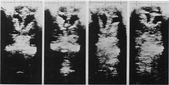
Two cases are described of a previously unrecognised sequel of posthaemorrhagic ventricular dilatation. The first case documents freely mobile blood clots within the lateral ventricular system, the second variable asymmetry in the size of the dilated lateral ventricle. The unilateral ventricular dilatation depended on which side the infant was lying, the dependent ventricle being considerably larger than the upper one within 4 hours of head turning. Each of these conditions spontaneously resolved with no specific treatment.

摘要

本文描述了两例出血后心室扩张先前未被认识到的后遗症。第一例记录了侧脑室系统内可自由移动的血凝块,第二例记录了扩张的侧脑室大小存在可变不对称性。单侧脑室扩张取决于婴儿的卧位,在转头后4小时内,受压侧脑室比上方侧脑室大得多。这些情况均未经特殊治疗而自行缓解。

https://cdn.ncbi.nlm.nih.gov/pmc/blobs/daae/1627524/be1211333319/archdisch00761-0077-b.jpg
https://cdn.ncbi.nlm.nih.gov/pmc/blobs/daae/1627524/716737395073/archdisch00761-0077-a.jpg
https://cdn.ncbi.nlm.nih.gov/pmc/blobs/daae/1627524/be1211333319/archdisch00761-0077-b.jpg
https://cdn.ncbi.nlm.nih.gov/pmc/blobs/daae/1627524/716737395073/archdisch00761-0077-a.jpg
https://cdn.ncbi.nlm.nih.gov/pmc/blobs/daae/1627524/be1211333319/archdisch00761-0077-b.jpg

相似文献

1
Elusive blood clots and fluctuating ventricular dilatation after neonatal intraventricular haemorrhage.新生儿脑室内出血后难以捉摸的血凝块和波动的脑室扩张。
Arch Dis Child. 1982 Feb;57(2):158-60. doi: 10.1136/adc.57.2.158.
2
Neonatal cerebral pathology diagnosed by ultrasound.
Clin Perinatol. 1985 Feb;12(1):195-218.
3
The occipital horn of the lateral ventricles in premature infants. An ultrasonographic study.
Eur J Radiol. 1983 May;3(2):148-50.
4
Neurosonographic identification of ventricular asymmetry in premature infants.
Clin Radiol. 1984 Jan;35(1):29-31. doi: 10.1016/s0009-9260(84)80225-1.
5
Neonatal intracranial hemorrhage and complicating hydrocephalus.新生儿颅内出血及并发脑积水。
J Clin Ultrasound. 1984 Jun;12(5):261-6. doi: 10.1002/jcu.1870120506.
6
Serial lumbar tapping to prevent posthaemorrhagic hydrocephalus after intracranial haemorrhage in preterm infants.连续腰椎穿刺预防早产儿颅内出血后出血后脑积水
Wien Klin Wochenschr. 1998 Oct 2;110(18):631-4.
7
Ultrasonic evaluation of neonatal intracranial hemorrhage and its complications.
Radiology. 1981 Jun;139(3):677-85. doi: 10.1148/radiology.139.3.7232735.
8
Real-time ultrasonography. Its use in the evaluation of neonatal intracranial hemorrhage and posthemorrhagic hydrocephalus.
Am J Dis Child. 1981 Apr;135(4):319-21.
9
Posthaemorrhagic ventricular dilatation.出血后脑室扩张
Arch Dis Child Fetal Neonatal Ed. 2002 Mar;86(2):F72-4. doi: 10.1136/fn.86.2.f72.
10
The relationship between head circumference and the development of communicating hydrocephalus in infants following intraventricular hemmorrhage.婴儿脑室内出血后头围与交通性脑积水发展之间的关系。
Pediatrics. 1975 Jul;56(1):74-7.

引用本文的文献

1
Post-Hemorrhagic Hydrocephalus and Outcomes Amongst Neonates With Intraventricular Hemorrhage: A Systematic Review and Pooled Analysis.脑室出血新生儿的出血后脑积水及其预后:一项系统评价和汇总分析
Cureus. 2021 Oct 18;13(10):e18877. doi: 10.7759/cureus.18877. eCollection 2021 Oct.
2
Neuroendoscopic fenestration of the foramen of Monro without septostomy for unilateral hydrocephalus following neonatal intraventricular hemorrhage.
Childs Nerv Syst. 2011 Mar;27(3):473-8. doi: 10.1007/s00381-010-1272-1. Epub 2010 Sep 2.
3
Neurosonography of hydrocephalus in infants.
Neuroradiology. 1986;28(5-6):452-62. doi: 10.1007/BF00344099.
4

本文引用的文献

1
A longitudinal study of post-haemorrhagic ventricular dilatation in the newborn.新生儿出血后脑室扩张的纵向研究。
Arch Dis Child. 1981 Dec;56(12):905-10. doi: 10.1136/adc.56.12.905.
2
Measurement of the growth of the lateral ventricles in preterm infants with real-time ultrasound.实时超声测量早产儿侧脑室的生长情况。
Arch Dis Child. 1981 Dec;56(12):900-4. doi: 10.1136/adc.56.12.900.
3
Cerebral structure and intraventricular haemorrhage in the neonate: a real-time ultrasound study.新生儿脑结构与脑室内出血:一项实时超声研究
Changes in the lateral ventricle with the head position: ultrasonographic observation.
Neuroradiology. 1988;30(4):315-8. doi: 10.1007/BF00328182.
Arch Dis Child. 1981 Jun;56(6):416-24. doi: 10.1136/adc.56.6.416.
4
Early increase in intracranial pressure in preterm infants.早产儿颅内压早期升高
Pediatrics. 1978 Jun;61(6):904-7.
5
Intraventricular hemorrhage and hydrocephalus in premature newborns: a prospective study with CT.早产儿脑室内出血与脑积水:一项CT前瞻性研究
AJR Am J Roentgenol. 1979 Apr;132(4):631-5. doi: 10.2214/ajr.132.4.631.
6
Ultrasound detection of brain damage in preterm infants.
Lancet. 1979 Jun 16;1(8129):1261-4. doi: 10.1016/s0140-6736(79)92227-x.