Rootman J, Goldberg C, Robertson W
Br J Ophthalmol. 1982 Mar;66(3):194-204. doi: 10.1136/bjo.66.3.194.
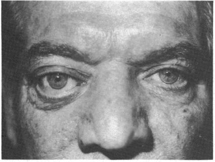
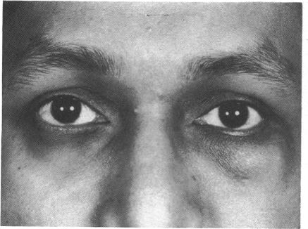
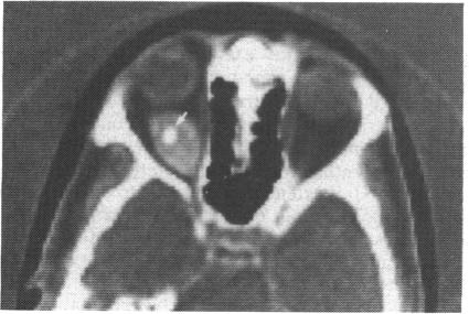
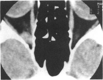
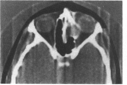
Seven histologically proved cases of primary orbital schwannoma have been seen at the University of British Columbia Orbital Clinic between September 1976 and December 1980. We describe here their varied clinical presentations, preoperative investigations, operative findings, and appearances on light and electron microscopy. Although no single feature is pathognomonic, a multiplicity of clinical, radiographic, and surgical features point to this lesion. Of preoperative investigations the computed tomography scan was the most helpful, especially in localising the lesion. Of the 7, 4 were intraconal and 3 were extraconal. The surgical approach was dictated by tumour site and included anterior, lateral, and panoramic orbitotomies. At surgery the nerve of origin of 4 of the tumours was identified. All tumours were excised totally or subtotally. There has been no recurrence to date.
1976年9月至1980年12月期间,不列颠哥伦比亚大学眼眶诊所共诊治了7例经组织学证实的原发性眼眶神经鞘瘤。在此,我们描述其多样的临床表现、术前检查、手术所见以及光镜和电镜下的表现。尽管没有单一特征具有确诊意义,但多种临床、影像学和手术特征指向该病变。术前检查中,计算机断层扫描最有帮助,尤其是在病变定位方面。7例中,4例位于肌锥内,3例位于肌锥外。手术入路取决于肿瘤部位,包括前路、外侧和眶周切开术。手术中确定了4例肿瘤的起源神经。所有肿瘤均被完全或次全切除。迄今为止,无复发情况。