• 文献检索
  • 文档翻译
  • 深度研究
  • 学术资讯
  • Suppr Zotero 插件Zotero 插件
  • 邀请有礼
  • 套餐&价格
  • 历史记录
应用&插件
Suppr Zotero 插件Zotero 插件浏览器插件Mac 客户端Windows 客户端微信小程序
定价
高级版会员购买积分包购买API积分包
服务
文献检索文档翻译深度研究API 文档MCP 服务
关于我们
关于 Suppr公司介绍联系我们用户协议隐私条款
关注我们

Suppr 超能文献

核心技术专利:CN118964589B侵权必究
粤ICP备2023148730 号-1Suppr @ 2026

文献检索

告别复杂PubMed语法,用中文像聊天一样搜索,搜遍4000万医学文献。AI智能推荐,让科研检索更轻松。

立即免费搜索

文件翻译

保留排版,准确专业,支持PDF/Word/PPT等文件格式,支持 12+语言互译。

免费翻译文档

深度研究

AI帮你快速写综述,25分钟生成高质量综述,智能提取关键信息,辅助科研写作。

立即免费体验

腰椎关节突关节的半月板。

The menisci of the lumbar zygapophysial joints.

作者信息

Engel R, Bogduk N

出版信息

J Anat. 1982 Dec;135(Pt 4):795-809.

PMID:7183677
原文链接:https://pmc.ncbi.nlm.nih.gov/articles/PMC1169447/
Abstract

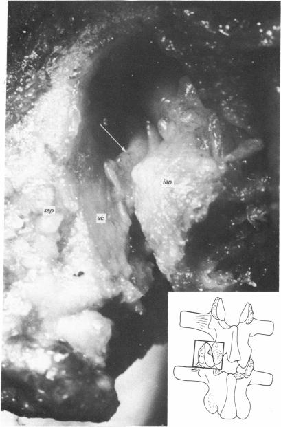
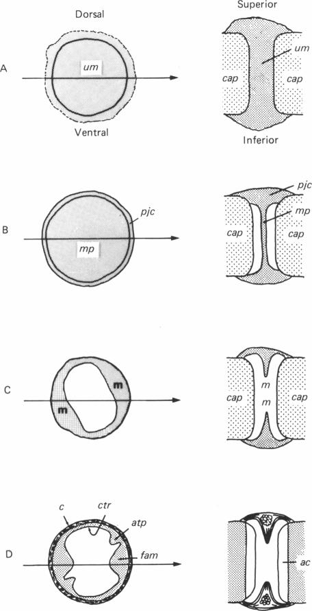
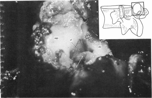
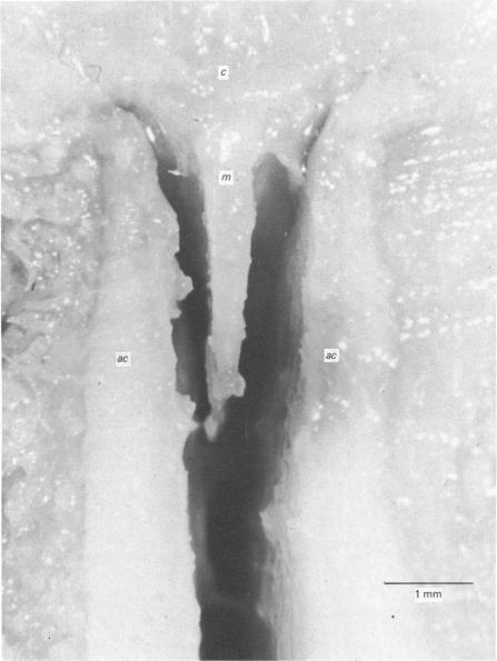
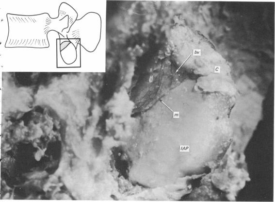
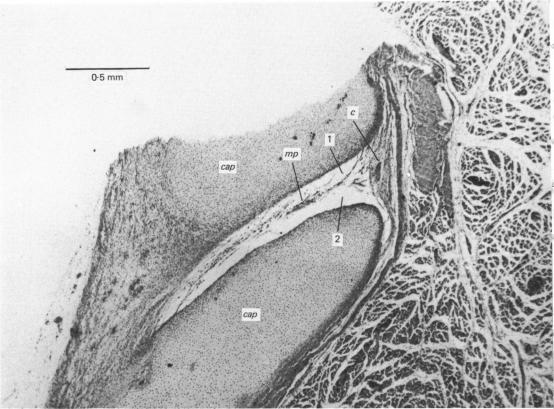
In a study of 82 lumbar zygapophysial joints three types of intra-articular structures were identified. They were adipose tissue pads and fibro-adipose meniscoids, both located at the superior and inferior poles of the joint, and connective tissue rims, located along the dorsal and ventral margins. Every lumbar zygapophysial joint contained at least one of these structures and 47 contained more than one type. Connective tissue rims are short, central projections of the joint capsule and do not enter between the articular surfaces. Adipose tissue pads are covered by synovium and fill the subcapsular space at the superoventral and inferodorsal poles of the joint. Fibro-adipose meniscoids, also covered by synovium, project from the joint capsule at the superior and inferior poles and enter between the articular surfaces. Adipose tissue pads and fibro-adipose meniscoids are probably derived from a common primitive mesenchymal meniscus which primarily differentiates into a fatty structure. The fibrous component of fibro-adipose meniscoids then secondarily develops as a result of compression of the tip of the fatty structure between the articular surfaces. The function of these intra-articular structures is not evident but may be related to protection of the articular processes as they subluxate during flexion and extension. Meniscus entrapment in the lumbar zygapophysial joints has been proposed as a cause of acute locked back, but the present morphological data are inconsistent with this view.

摘要

在一项对82个腰椎关节突关节的研究中,识别出了三种关节内结构。它们是脂肪组织垫和纤维脂肪半月板样结构,均位于关节的上极和下极,以及结缔组织边缘,位于背侧和腹侧边缘。每个腰椎关节突关节至少包含这些结构中的一种,47个关节包含不止一种类型。结缔组织边缘是关节囊的短的中央突起,不进入关节面之间。脂肪组织垫被滑膜覆盖,填充关节上腹部和下背部极的囊下间隙。纤维脂肪半月板样结构也被滑膜覆盖,从关节囊的上极和下极突出并进入关节面之间。脂肪组织垫和纤维脂肪半月板样结构可能源自共同的原始间充质半月板,其主要分化为脂肪结构。纤维脂肪半月板样结构的纤维成分随后由于脂肪结构的尖端在关节面之间受到挤压而继发形成。这些关节内结构的功能尚不清楚,但可能与在屈伸过程中关节突半脱位时对其的保护有关。有人提出腰椎关节突关节中的半月板嵌顿是急性腰背痛的一个原因,但目前的形态学数据与这一观点不一致。

https://cdn.ncbi.nlm.nih.gov/pmc/blobs/b501/1169447/be149ae23a99/janat00216-0135-a.jpg
https://cdn.ncbi.nlm.nih.gov/pmc/blobs/b501/1169447/3a0f276380be/janat00216-0127-a.jpg
https://cdn.ncbi.nlm.nih.gov/pmc/blobs/b501/1169447/1ed640c2addb/janat00216-0128-a.jpg
https://cdn.ncbi.nlm.nih.gov/pmc/blobs/b501/1169447/371f07aa8187/janat00216-0129-a.jpg
https://cdn.ncbi.nlm.nih.gov/pmc/blobs/b501/1169447/d42bf6b79b72/janat00216-0130-a.jpg
https://cdn.ncbi.nlm.nih.gov/pmc/blobs/b501/1169447/965e60d0fa4e/janat00216-0132-a.jpg
https://cdn.ncbi.nlm.nih.gov/pmc/blobs/b501/1169447/86026e34358d/janat00216-0133-a.jpg
https://cdn.ncbi.nlm.nih.gov/pmc/blobs/b501/1169447/52eb424572d0/janat00216-0134-a.jpg
https://cdn.ncbi.nlm.nih.gov/pmc/blobs/b501/1169447/be149ae23a99/janat00216-0135-a.jpg

相似文献

1
The menisci of the lumbar zygapophysial joints.腰椎关节突关节的半月板。
J Anat. 1982 Dec;135(Pt 4):795-809.
2
The menisci of the lumbar zygapophyseal joints. A review of their anatomy and clinical significance.
Spine (Phila Pa 1976). 1984 Jul-Aug;9(5):454-60. doi: 10.1097/00007632-198407000-00006.
3
[Meniscoids of the intervertebral joints].[椎间关节的半月板样结构]
Acta Chir Orthop Traumatol Cech. 2002;69(3):149-57.
4
Intra-articular inclusions of the cervical synovial joints.颈椎滑膜关节的关节内包涵体。
Br J Rheumatol. 1993 Aug;32(8):705-10. doi: 10.1093/rheumatology/32.8.705.
5
Intra-articular meniscoid folds in thoracic zygapophysial joints.胸椎间关节内的关节内半月板样皱襞。
Spine (Phila Pa 1976). 2010 Mar 15;35(6):E191-7. doi: 10.1097/BRS.0b013e3181c9b053.
6
Lumbar facet joint capsule: appearance at MR imaging and CT.腰椎小关节囊:磁共振成像和计算机断层扫描表现
Radiology. 1990 Nov;177(2):415-20. doi: 10.1148/radiology.177.2.2217778.
7
Lumbar facet joint fat pads: their normal anatomy and their appearance when enlarged.腰椎小关节脂肪垫:其正常解剖结构及增大时的表现。
Neuroradiology. 1991;33(1):38-42. doi: 10.1007/BF00593331.
8
Intra-articular synovial folds of thoracolumbar junction zygapophyseal joints.胸腰段关节突关节的关节内滑膜皱襞
Anat Rec. 1990 Feb;226(2):147-52. doi: 10.1002/ar.1092260204.
9
The anatomy and morphometry of cervical zygapophyseal joint meniscoids.颈椎关节突关节半月板的解剖学与形态测量学
Surg Radiol Anat. 2015 Sep;37(7):799-807. doi: 10.1007/s00276-014-1406-3. Epub 2014 Dec 20.
10
Intra-articular inclusions of the elbow joint complex.
Clin Anat. 2007 Aug;20(6):668-76. doi: 10.1002/ca.20467.

引用本文的文献

1
[Lumbar Facet Joint Injection: A Review of Efficacy and Safety].[腰椎小关节注射:疗效与安全性综述]
J Korean Soc Radiol. 2024 Jan;85(1):54-76. doi: 10.3348/jksr.2023.0146. Epub 2024 Jan 26.
2
Facet Joint Syndrome: Pathophysiology, Diagnosis, and Treatment.小关节综合征:病理生理学、诊断与治疗
J Pain Res. 2022 Nov 30;15:3689-3710. doi: 10.2147/JPR.S389602. eCollection 2022.
3
Facet joint injections for management of low back pain: a clinically focused review.用于管理下腰痛的小关节注射:一项临床重点综述。

本文引用的文献

1
The development of joints.关节的发育。
J Anat. 1947 Jan;81(Pt 1):33-55.
2
The development of synovial joints.滑膜关节的发育。
J Anat. 1940 Jan;74(Pt 2):277-83.
3
The morphology of the lumbar synovial interveertebral joints.腰椎滑膜关节的形态学
Anesth Pain Med (Seoul). 2020 Jan 31;15(1):8-18. doi: 10.17085/apm.2020.15.1.8.
4
A temporary disc-like structure at the median atlanto-axial joint in human fetuses.人类胎儿寰枢正中关节处的一种临时性盘状结构。
Anat Cell Biol. 2019 Dec;52(4):436-442. doi: 10.5115/acb.19.128. Epub 2019 Dec 31.
5
Anatomical etiology of "pseudo-sciatica" from superior cluneal nerve entrapment: a laboratory investigation.臀上皮神经卡压所致“假性坐骨神经痛”的解剖学病因:一项实验室研究
J Pain Res. 2017 Nov 1;10:2539-2545. doi: 10.2147/JPR.S142115. eCollection 2017.
6
Expression of adiponectin in the subchondral bone of lumbar facet joints with different degrees of degeneration.脂联素在不同退变程度腰椎小关节软骨下骨中的表达。
BMC Musculoskelet Disord. 2017 Nov 3;18(1):427. doi: 10.1186/s12891-017-1786-8.
7
The use of functional and traditional mobilization interventions in a patient with chronic thoracic pain: a case report.功能性和传统松动干预措施在一名慢性胸痛患者中的应用:病例报告
J Man Manip Ther. 2013 Aug;21(3):134-41. doi: 10.1179/2042618612Y.0000000024.
8
Magnetic resonance imaging zygapophyseal joint space changes (gapping) in low back pain patients following spinal manipulation and side-posture positioning: a randomized controlled mechanisms trial with blinding.脊柱推拿和侧卧位后腰痛患者的磁共振成像关节突关节间隙变化(间隙增宽):一项采用盲法的随机对照机制试验
J Manipulative Physiol Ther. 2013 May;36(4):203-17. doi: 10.1016/j.jmpt.2013.04.003. Epub 2013 May 3.
9
Spinal facet joint biomechanics and mechanotransduction in normal, injury and degenerative conditions.正常、损伤及退变情况下的脊柱小关节生物力学与机械转导
J Biomech Eng. 2011 Jul;133(7):071010. doi: 10.1115/1.4004493.
10
Morphometry of the synovial folds of the lateral atlanto-axial joints: the anatomical basis for understanding their potential role in neck pain.寰枢外侧关节滑膜皱襞的形态测量学:理解其在颈部疼痛中潜在作用的解剖学基础。
Surg Radiol Anat. 2012 Mar;34(2):115-24. doi: 10.1007/s00276-011-0834-6. Epub 2011 Jun 12.
Acta Morphol Neerl Scand. 1962;4:299-319.
4
[ORIGIN, STRUCTURE AND FUNCTION OF MENISCOID STRUCTURES IN THE CERVICAL VERTEBRAL JOINTS].
Z Orthop Ihre Grenzgeb. 1963 Dec;98:1-14.
5
Anatomico-roentgenographic studies of the posterior spinal articulations.脊柱后关节的解剖X线研究
Am J Roentgenol Radium Ther Nucl Med. 1961 Aug;86:270-6.
6
[Anatomy of the vertebral joint].
Arch Orthop Unfallchir. 1958;50(3):222-34. doi: 10.1007/BF00416096.
7
[Research on the articulations between the articular processes of human vertebrae. I. Several little noted and delicate arrangements of the joint apparatus].[人类椎骨关节突之间关节的研究。I. 关节装置的一些鲜为人知且精细的结构]
Arch Ital Anat Embriol. 1956;61(1):1-16.
8
[Mortality in puppies caused by myocarditis].[幼犬心肌炎所致死亡]
Tijdschr Diergeneeskd. 1979 Aug 15;104(15-16):634-5.