• 文献检索
  • 文档翻译
  • 深度研究
  • 学术资讯
  • Suppr Zotero 插件Zotero 插件
  • 邀请有礼
  • 套餐&价格
  • 历史记录
应用&插件
Suppr Zotero 插件Zotero 插件浏览器插件Mac 客户端Windows 客户端微信小程序
定价
高级版会员购买积分包购买API积分包
服务
文献检索文档翻译深度研究API 文档MCP 服务
关于我们
关于 Suppr公司介绍联系我们用户协议隐私条款
关注我们

Suppr 超能文献

核心技术专利:CN118964589B侵权必究
粤ICP备2023148730 号-1Suppr @ 2026

文献检索

告别复杂PubMed语法,用中文像聊天一样搜索,搜遍4000万医学文献。AI智能推荐,让科研检索更轻松。

立即免费搜索

文件翻译

保留排版,准确专业,支持PDF/Word/PPT等文件格式,支持 12+语言互译。

免费翻译文档

深度研究

AI帮你快速写综述,25分钟生成高质量综述,智能提取关键信息,辅助科研写作。

立即免费体验

小鼠脊髓中央管室管膜发育的电子显微镜研究。

An electron microscopic study of the development of the ependyma of the central canal of the mouse spinal cord.

作者信息

Sturrock R R

出版信息

J Anat. 1981 Jan;132(Pt 1):119-36.

PMID:7275786
原文链接:https://pmc.ncbi.nlm.nih.gov/articles/PMC1233400/
Abstract

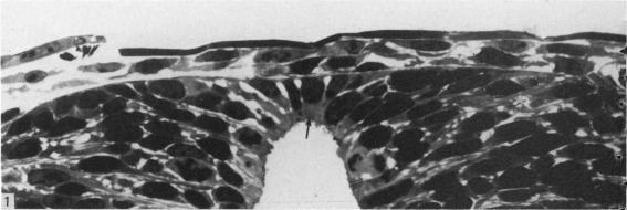
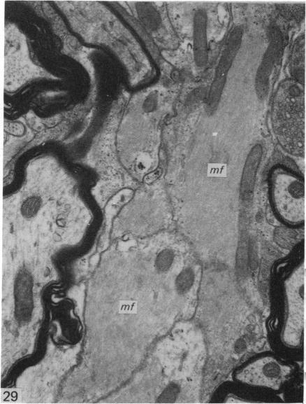
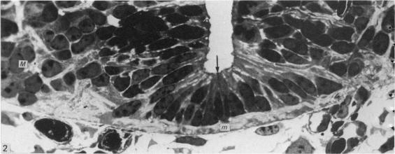
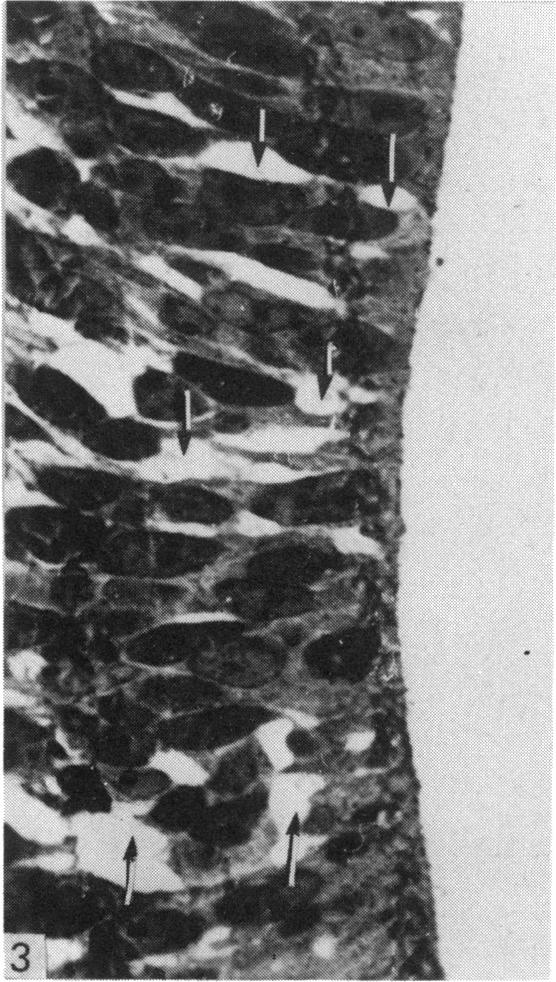
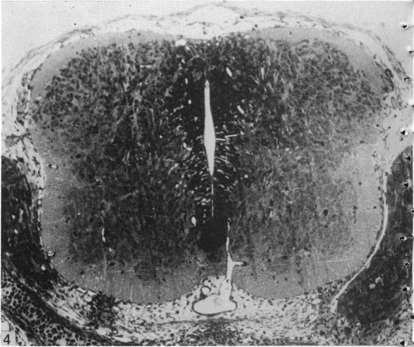
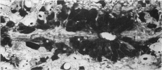
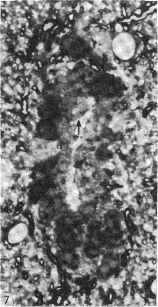
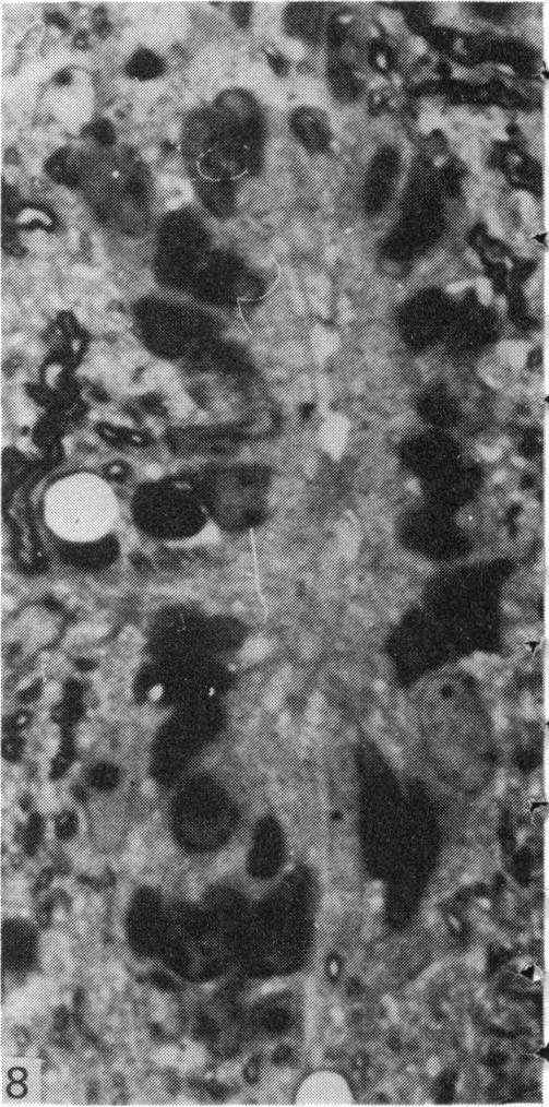
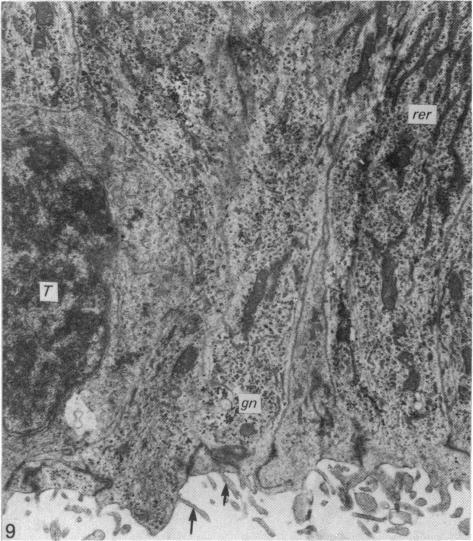
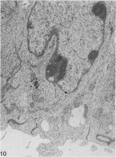
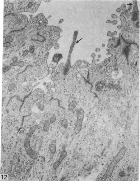
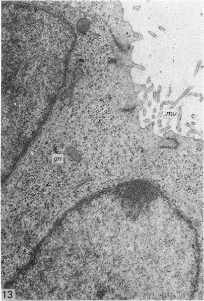
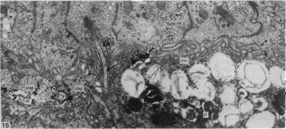
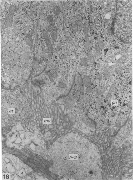
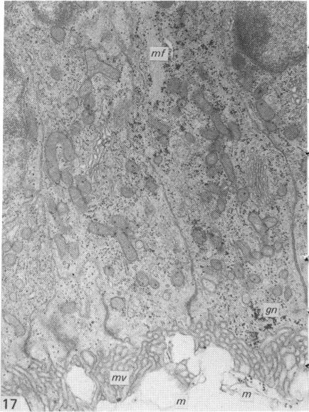
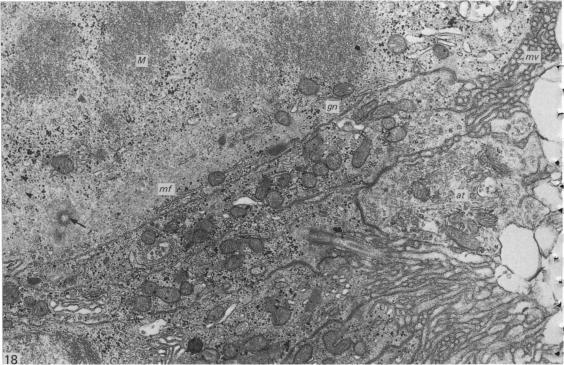
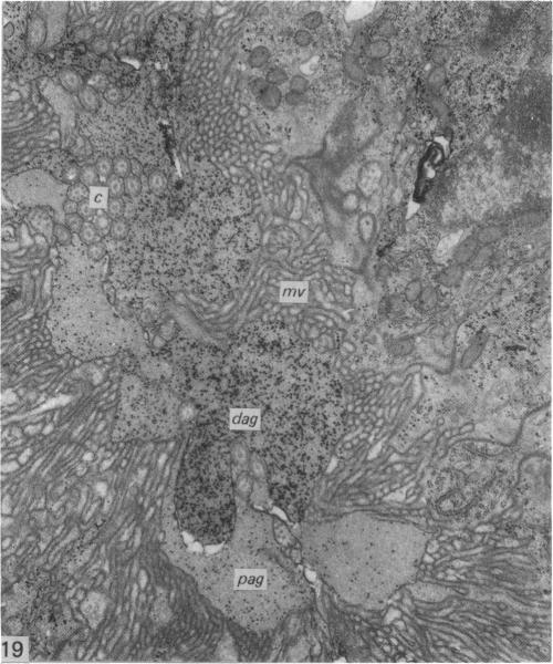
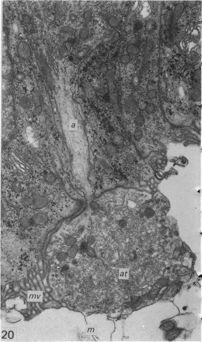
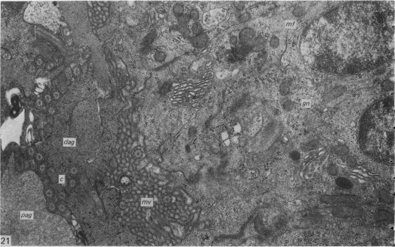
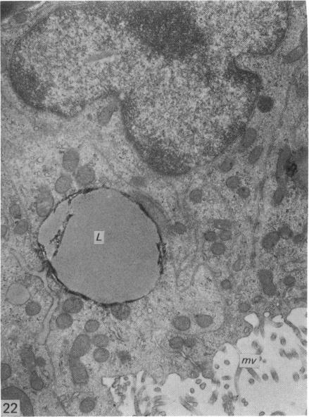
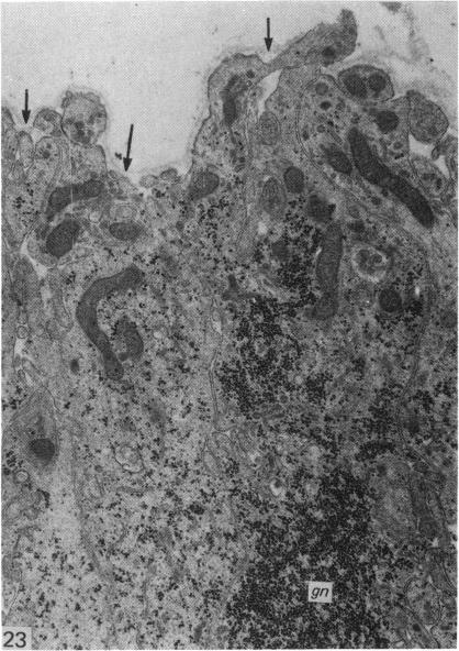
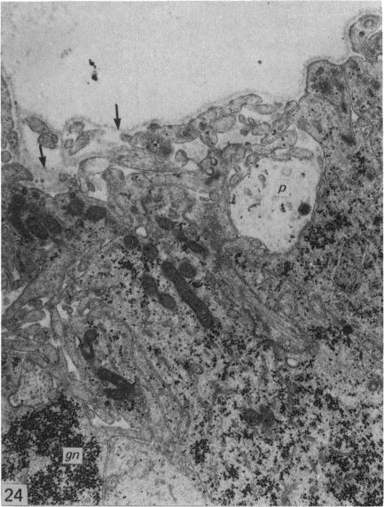
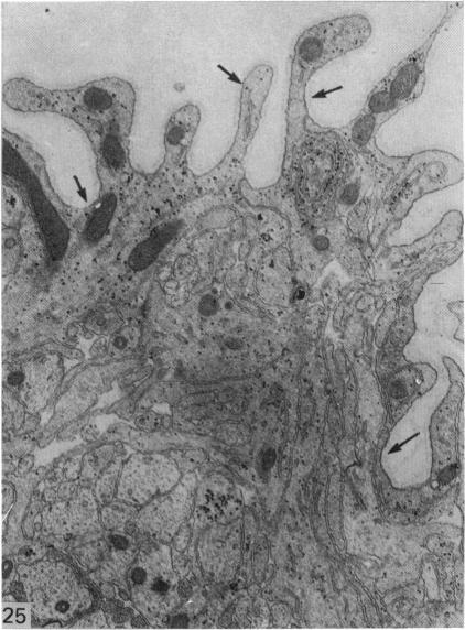
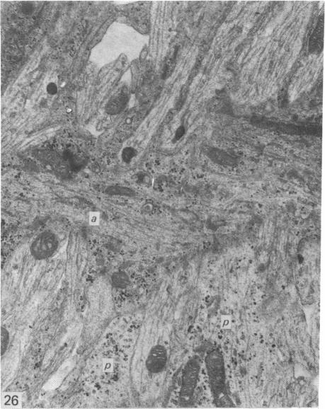
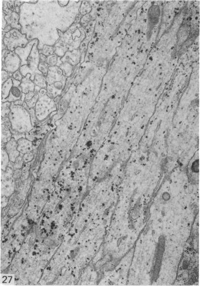
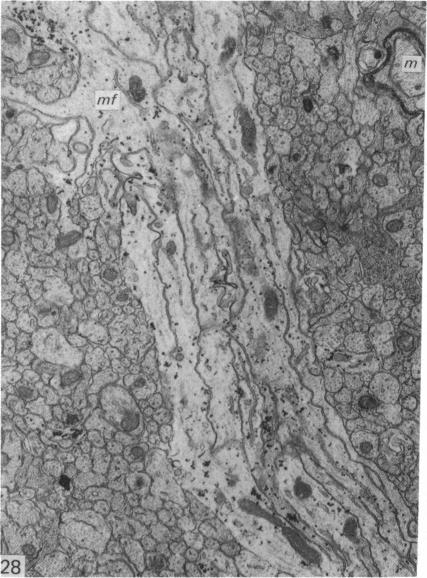
The central canal of the adult mouse spinal cord is lined for most of its extent by ependymal cells which are rich in microfilaments and whose apical surface is covered with matted, broad microvilli. The canal itself is filled with amorphous material containing glycogen granules. Two forms of this material are present, a dark form rich in glycogen, and a light form containing a few glycogen granules. Each type appears to be surrounded by a membrane. The upper cervical region, however, has a large empty lumen and the ependymal cells in this region have only scattered, narrow microvilli. During development, the floor and roof plates are at first composed largely of ependymoglial cells, unlike the lateral walls, where undifferentiated neuroepithelial cells predominate. By E15 few undifferentiated neuroepithelial cells remain. At E17 the morphology of the ependymal cells changes. Their apical surface becomes covered with matted, club-shaped microvilli and the central canal is filled with glycogen-containing material. By P5 microfibrils are present in large bundles in the ependymal cells. The piaglial surface opposite the roof and floor plates has finger-like projections unique to these regions and these persist at the surface of the dorsal median septum until myelination is well advanced after P5. The fibres forming the dorsal median septum are at first pale processes containing scattered glycogen granules and microtubules. By P5 microfibrils are present and at P150 the processes are packed with masses of microfibrils.

摘要

成年小鼠脊髓的中央管大部分长度由室管膜细胞衬里,这些细胞富含微丝,其顶端表面覆盖着交织在一起的宽微绒毛。管腔内充满含有糖原颗粒的无定形物质。这种物质有两种形式,一种深色形式富含糖原,另一种浅色形式含有少量糖原颗粒。每种类型似乎都被一层膜包围着。然而,上颈段有一个大的空管腔,该区域的室管膜细胞只有散在的窄微绒毛。在发育过程中,底板和顶板最初主要由室管膜胶质细胞组成,这与侧壁不同,侧壁中未分化的神经上皮细胞占主导。到胚胎第15天(E15),很少有未分化的神经上皮细胞留存。在胚胎第17天(E17),室管膜细胞的形态发生变化。它们的顶端表面被交织在一起的棒状微绒毛覆盖,中央管充满含糖原物质。到出生后第5天(P5),室管膜细胞中有大量成束的微原纤维。与顶板和底板相对的软膜表面有这些区域特有的指状突起,这些突起在背正中隔表面持续存在,直到出生后第5天(P5)髓鞘形成进展良好。形成背正中隔的纤维最初是浅色突起,含有散在的糖原颗粒和微管。到出生后第5天(P5)出现微原纤维,到出生后第150天(P150),突起中充满大量微原纤维。

https://cdn.ncbi.nlm.nih.gov/pmc/blobs/af69/1233400/5aba120e86a9/janat00225-0137-d.jpg
https://cdn.ncbi.nlm.nih.gov/pmc/blobs/af69/1233400/60b27900248a/janat00225-0123-a.jpg
https://cdn.ncbi.nlm.nih.gov/pmc/blobs/af69/1233400/2e6764eed0bb/janat00225-0123-b.jpg
https://cdn.ncbi.nlm.nih.gov/pmc/blobs/af69/1233400/0a91ed4e71bb/janat00225-0123-c.jpg
https://cdn.ncbi.nlm.nih.gov/pmc/blobs/af69/1233400/6822c9401082/janat00225-0123-d.jpg
https://cdn.ncbi.nlm.nih.gov/pmc/blobs/af69/1233400/618cd063c5bd/janat00225-0125-a.jpg
https://cdn.ncbi.nlm.nih.gov/pmc/blobs/af69/1233400/2f010a7adc28/janat00225-0125-b.jpg
https://cdn.ncbi.nlm.nih.gov/pmc/blobs/af69/1233400/a730da4b1fee/janat00225-0125-c.jpg
https://cdn.ncbi.nlm.nih.gov/pmc/blobs/af69/1233400/d2e579fbcb52/janat00225-0125-d.jpg
https://cdn.ncbi.nlm.nih.gov/pmc/blobs/af69/1233400/85f97b309d63/janat00225-0127-a.jpg
https://cdn.ncbi.nlm.nih.gov/pmc/blobs/af69/1233400/567c2e0dc03e/janat00225-0127-b.jpg
https://cdn.ncbi.nlm.nih.gov/pmc/blobs/af69/1233400/1cb27ce522f8/janat00225-0127-c.jpg
https://cdn.ncbi.nlm.nih.gov/pmc/blobs/af69/1233400/b3d1dbe9ac6b/janat00225-0127-d.jpg
https://cdn.ncbi.nlm.nih.gov/pmc/blobs/af69/1233400/ac1af9f7dd88/janat00225-0129-a.jpg
https://cdn.ncbi.nlm.nih.gov/pmc/blobs/af69/1233400/d2765e0d4dce/janat00225-0129-b.jpg
https://cdn.ncbi.nlm.nih.gov/pmc/blobs/af69/1233400/f6b00f43eb25/janat00225-0129-c.jpg
https://cdn.ncbi.nlm.nih.gov/pmc/blobs/af69/1233400/f8e29e1a05b8/janat00225-0131-a.jpg
https://cdn.ncbi.nlm.nih.gov/pmc/blobs/af69/1233400/3f573bf67385/janat00225-0131-b.jpg
https://cdn.ncbi.nlm.nih.gov/pmc/blobs/af69/1233400/6b0e26ff57b5/janat00225-0131-c.jpg
https://cdn.ncbi.nlm.nih.gov/pmc/blobs/af69/1233400/6803e6cef321/janat00225-0133-a.jpg
https://cdn.ncbi.nlm.nih.gov/pmc/blobs/af69/1233400/d6c82d50ba39/janat00225-0133-b.jpg
https://cdn.ncbi.nlm.nih.gov/pmc/blobs/af69/1233400/962bee53f00a/janat00225-0133-c.jpg
https://cdn.ncbi.nlm.nih.gov/pmc/blobs/af69/1233400/3c53404b5e07/janat00225-0135-a.jpg
https://cdn.ncbi.nlm.nih.gov/pmc/blobs/af69/1233400/2ead765e01a4/janat00225-0135-b.jpg
https://cdn.ncbi.nlm.nih.gov/pmc/blobs/af69/1233400/9fa5a41b14f2/janat00225-0135-c.jpg
https://cdn.ncbi.nlm.nih.gov/pmc/blobs/af69/1233400/49b756903fe5/janat00225-0135-d.jpg
https://cdn.ncbi.nlm.nih.gov/pmc/blobs/af69/1233400/8b070d984910/janat00225-0137-a.jpg
https://cdn.ncbi.nlm.nih.gov/pmc/blobs/af69/1233400/89d6e27fb953/janat00225-0137-b.jpg
https://cdn.ncbi.nlm.nih.gov/pmc/blobs/af69/1233400/0a6750f94d36/janat00225-0137-c.jpg
https://cdn.ncbi.nlm.nih.gov/pmc/blobs/af69/1233400/5aba120e86a9/janat00225-0137-d.jpg

相似文献

1
An electron microscopic study of the development of the ependyma of the central canal of the mouse spinal cord.小鼠脊髓中央管室管膜发育的电子显微镜研究。
J Anat. 1981 Jan;132(Pt 1):119-36.
2
[Comparative scanning and transmission electron microscopy studies of the ependyma of the central canal in the spinal cord of primates. I. Electron optical image of the ependyma in the central canal of the spinal cord of the callithrix monkey (Callithrix jacchus, Linné 1758)].[灵长类动物脊髓中央管室管膜的比较扫描和透射电子显微镜研究。I. 狨猴(Callithrix jacchus,林奈,1758年)脊髓中央管室管膜的电子光学图像]
Gegenbaurs Morphol Jahrb. 1986;132(4):535-54.
3
[Transmission electron microscopic studies of the organization of the ependymocytes in the central canal of the spinal cord of Cercopithecus nigroviridis (Pocock 1907), (Platyrrhina, Cercopithecoidea)].[黑绿猴(Pocock,1907)(阔鼻猴亚目,猕猴科)脊髓中央管室管膜细胞组织结构的透射电子显微镜研究]
Gegenbaurs Morphol Jahrb. 1988;134(5):625-35.
4
A comparison of the processes of ventricular coarctation and choroid and ependymal fusion in the mouse brain.小鼠脑室缩窄与脉络膜和室管膜融合过程的比较。
J Anat. 1979 Sep;129(Pt 2):235-42.
5
[The cerebrospinal fluid contact processes in the central canal of the spinal cord. A scanning and transmission electron microscopic study of the rabbit].[脊髓中央管中的脑脊液接触过程。对兔的扫描和透射电子显微镜研究]
Z Mikrosk Anat Forsch. 1976;90(1):1-15.
6
Formation of the central canal and dorsal glial septum in the spinal cord of the domestic cat.家猫脊髓中央管和背侧神经胶质隔的形成
J Anat. 1988 Aug;159:37-47.
7
Electron microscopic study of the progeny of ependymal stem cells in the normal and injured spinal cord.正常和损伤脊髓室管膜干细胞子代的电子显微镜研究
Surg Neurol. 2005;64 Suppl 2:S28-32. doi: 10.1016/j.surneu.2005.07.057.
8
The ependyma of the cat central canal, with particular reference to its mitochondria-containing bulbs.猫中央管的室管膜,特别涉及其含线粒体的球状体。
Scan Electron Microsc. 1985(Pt 1):231-8.
9
Fine structure of regenerated ependyma and spinal cord in Sternarchus albifrons.白边电鳗再生室管膜和脊髓的精细结构
Anat Rec. 1983 Jan;205(1):73-83. doi: 10.1002/ar.1092050110.
10
Ependyma of the central canal of the rat spinal cord: a light and transmission electron microscopic study.大鼠脊髓中央管室管膜:光镜与透射电镜研究
J Anat. 1987 Jun;152:55-70.

引用本文的文献

1
Wnt-dependent mechanism of the apical constriction of roof plate cells in developing mouse spinal cord.发育中小鼠脊髓顶板细胞顶端收缩的Wnt依赖机制。
Front Cell Dev Biol. 2025 Jul 7;13:1571770. doi: 10.3389/fcell.2025.1571770. eCollection 2025.
2
Morphological and Functional Changes of Roof Plate Cells in Spinal Cord Development.脊髓发育过程中顶板细胞的形态学和功能变化
J Dev Biol. 2021 Jul 30;9(3):30. doi: 10.3390/jdb9030030.
3
Crumbs2 mediates ventricular layer remodelling to form the spinal cord central canal.Crumb2 介导室管膜层重塑以形成脊髓中央管。

本文引用的文献

1
An electron microscope study of ependymal cells of the fetal, early postnatal and adult rabbit.对胎儿、出生后早期及成年家兔室管膜细胞的电子显微镜研究。
Z Zellforsch Mikrosk Anat. 1962;56:595-618. doi: 10.1007/BF00540584.
2
A morphological study of the mouse subependymal layer from embryonic life to old age.从小鼠胚胎期至老年期室管膜下层的形态学研究。
J Anat. 1980 Mar;130(Pt 2):391-415.
3
Fine structure of ependyma in spinal cord of human embryos.
Folia Morphol (Praha). 1967;15(1):68-78.
PLoS Biol. 2020 Mar 9;18(3):e3000470. doi: 10.1371/journal.pbio.3000470. eCollection 2020 Mar.
4
Multiple steps characterise ventricular layer attrition to form the ependymal cell lining of the adult mouse spinal cord central canal.心室层磨损形成成年小鼠脊髓中央管室管膜细胞 lining 需要经过多个步骤。
J Anat. 2020 Feb;236(2):334-350. doi: 10.1111/joa.13094. Epub 2019 Oct 31.
5
Roles of axon guidance molecules in neuronal wiring in the developing spinal cord.轴突导向分子在发育中脊髓神经元布线中的作用。
Nat Rev Neurosci. 2019 Jul;20(7):380-396. doi: 10.1038/s41583-019-0168-7.
6
Sensory and spinal inhibitory dorsal midline crossing is independent of Robo3.感觉性和脊髓抑制性背侧中线交叉独立于Robo3。
Front Neural Circuits. 2015 Jul 23;9:36. doi: 10.3389/fncir.2015.00036. eCollection 2015.
7
Stretching morphogenesis of the roof plate and formation of the central canal.脊索诱导神经管形成和室管膜的延伸。
PLoS One. 2013;8(2):e56219. doi: 10.1371/journal.pone.0056219. Epub 2013 Feb 7.
8
Role of radial glia in transformation of the primitive lumen to the central canal in the developing rat spinal cord.放射状胶质细胞在发育中的大鼠脊髓原始管腔向中央管转变中的作用。
Cell Mol Neurobiol. 2009 Sep;29(6-7):927-36. doi: 10.1007/s10571-009-9377-3. Epub 2009 Mar 17.
9
Reinforcement of cell junctions correlates with the absence of hair cell regeneration in mammals and its occurrence in birds.细胞连接的强化与哺乳动物中毛细胞再生的缺失及其在鸟类中的出现相关。
J Comp Neurol. 2008 Nov 20;511(3):396-414. doi: 10.1002/cne.21849.
10
Ultrastructural and cytochemical characterisation of the floor plate ependyma of the developing rat spinal cord.发育中大鼠脊髓室管膜底板的超微结构和细胞化学特征
J Anat. 1989 Aug;165:87-100.
4
Electron microscopic studies on Reissner's fiber and the ependymal cells in the spinal cord of the rat.
Z Zellforsch Mikrosk Anat. 1969;94(4):565-73. doi: 10.1007/BF00936062.
5
Electron microscope observations on the human foetal and embryonic spinal cord.对人类胎儿和胚胎脊髓的电子显微镜观察。
J Anat. 1969 May;104(Pt 3):435-53.
6
The neuroglia in the spinal cord of the newt, Triturus viridescens.绿螈(Triturus viridescens)脊髓中的神经胶质细胞。
J Comp Neurol. 1969 Jan;135(1):93-120. doi: 10.1002/cne.901350107.
7
[On the question of intraventricular neurosecretion. A nervous structure unknown until now in the IV ventricle of rabbits].[关于脑室内神经分泌的问题。家兔第四脑室内迄今未知的一种神经结构]
Z Zellforsch Mikrosk Anat. 1967;79(2):172-84.
8
[On the wall structure of the third ventricle in albino rats].[关于白化大鼠第三脑室的壁结构]
Histochemie. 1967;11(1):40-61. doi: 10.1007/BF00326611.
9
[Topographic distribution of supraependymal structures in the ventricles and central canal of the rabbit brain].
Z Zellforsch Mikrosk Anat. 1969 Sep 22;100(4):536-51.
10
Electron microscope observations upon the conus medullaris and filum terminale of human fetuses.人胎儿脊髓圆锥和终丝的电子显微镜观察。
J Anat. 1971 Nov;110(Pt 2):173-9.