• 文献检索
  • 文档翻译
  • 深度研究
  • 学术资讯
  • Suppr Zotero 插件Zotero 插件
  • 邀请有礼
  • 套餐&价格
  • 历史记录
应用&插件
Suppr Zotero 插件Zotero 插件浏览器插件Mac 客户端Windows 客户端微信小程序
定价
高级版会员购买积分包购买API积分包
服务
文献检索文档翻译深度研究API 文档MCP 服务
关于我们
关于 Suppr公司介绍联系我们用户协议隐私条款
关注我们

Suppr 超能文献

核心技术专利:CN118964589B侵权必究
粤ICP备2023148730 号-1Suppr @ 2026

文献检索

告别复杂PubMed语法,用中文像聊天一样搜索,搜遍4000万医学文献。AI智能推荐,让科研检索更轻松。

立即免费搜索

文件翻译

保留排版,准确专业,支持PDF/Word/PPT等文件格式,支持 12+语言互译。

免费翻译文档

深度研究

AI帮你快速写综述,25分钟生成高质量综述,智能提取关键信息,辅助科研写作。

立即免费体验

兔睾丸内收缩细胞的出生后发育与分化。

Postnatal development and differentiation of contractile cells within the rabbit testis.

作者信息

Leeson C R, Forman D E

出版信息

J Anat. 1981 Jun;132(Pt 4):491-511.

PMID:7298494
原文链接:https://pmc.ncbi.nlm.nih.gov/articles/PMC1233317/
Abstract

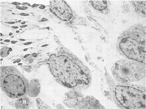
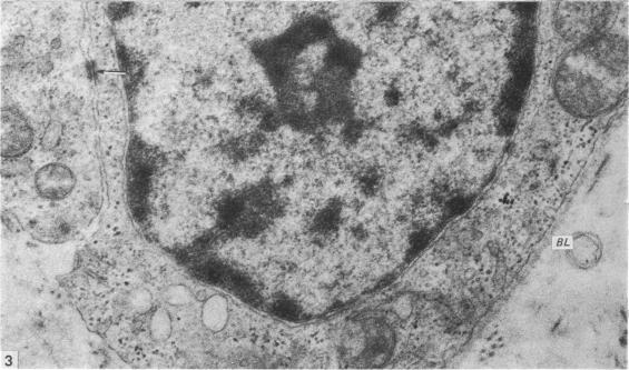
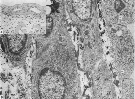
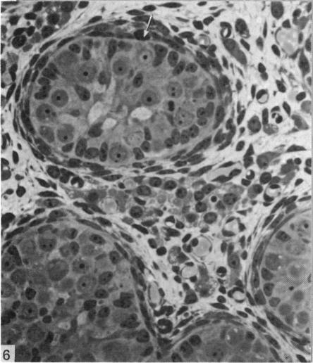
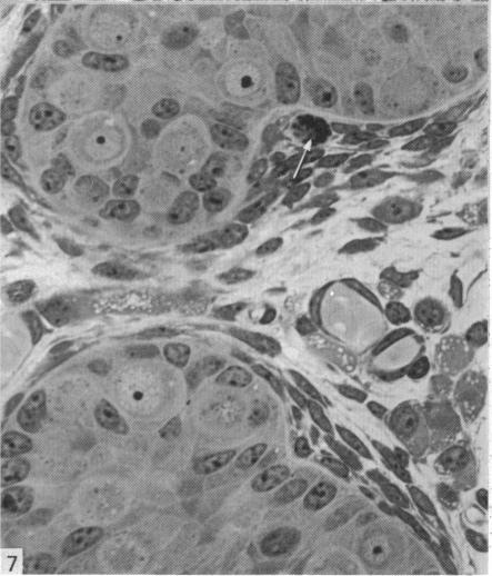
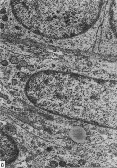
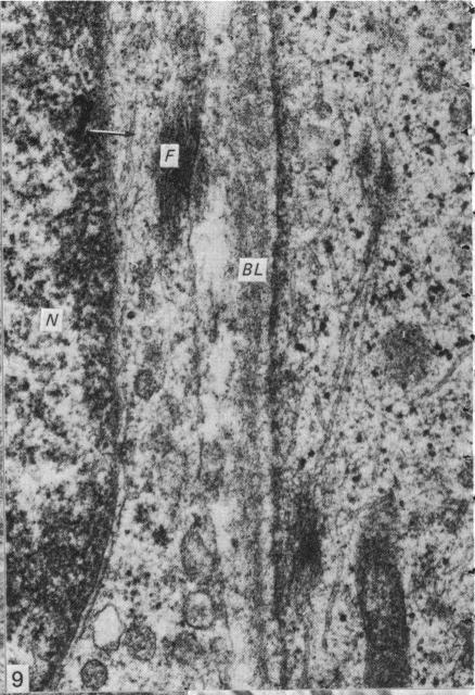
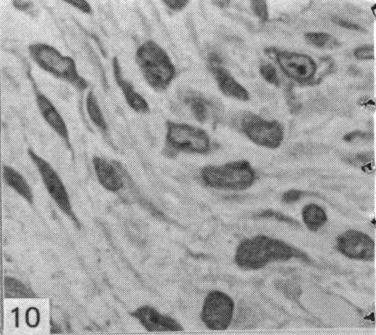
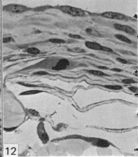
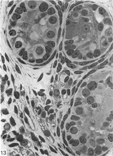
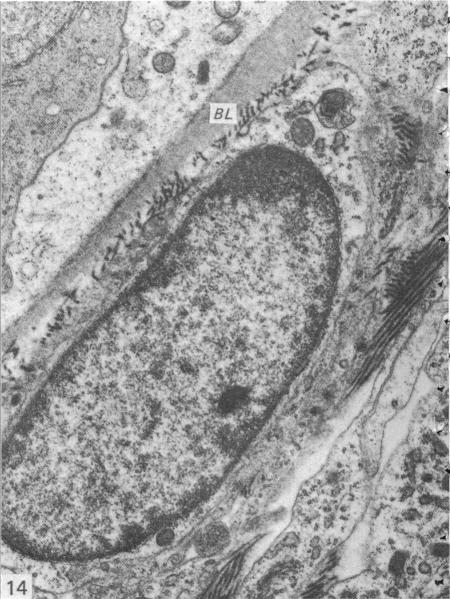
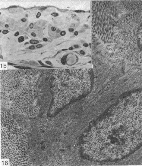
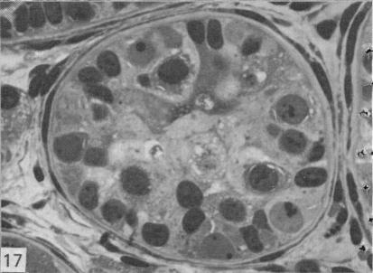
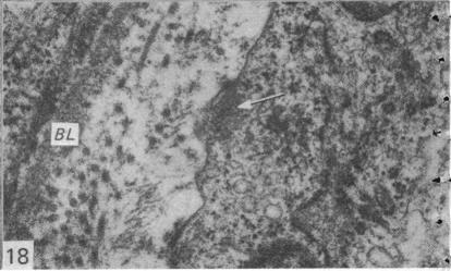
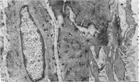
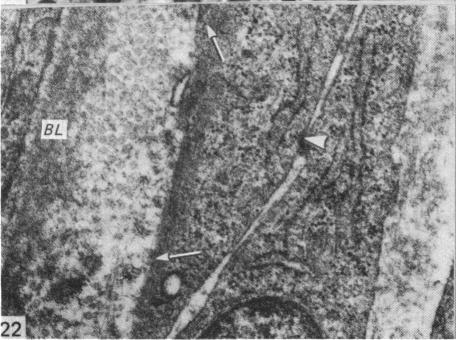
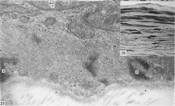
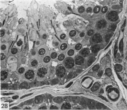
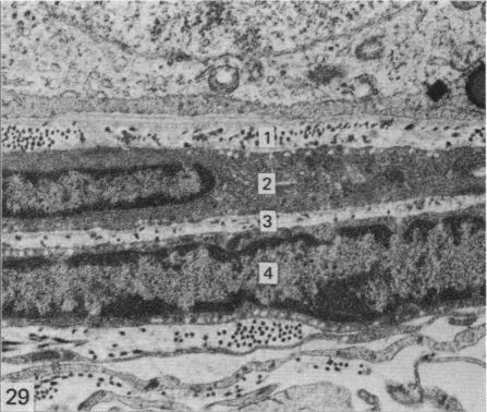
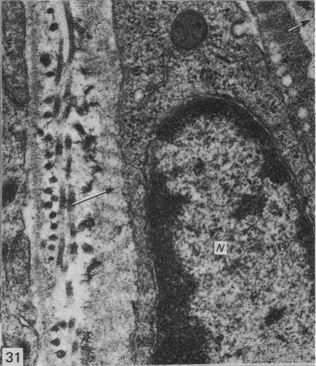
This study has been determined the postnatal development and differentiation of smooth muscle cells within the rabbit testicular capsule and within the peritubular tissue surrounding seminiferous tubules. Smooth muscle cells within the tunica albuginea are not identifiable at birth by light microscopy but by electron microscopy myocytes in early stages of development may be shown to be present. It is not until 42 to 49 days postnatum that smooth muscle cells can be identified by light microscopy. Differentiation of smooth muscle cells within the capsule is completed by 128 days postnatum. At this time, the muscle is arranged in two organized layers, a superficial layer of longitudinally oriented cells and a deeper layer of circularly arranged cells. At birth, the peritubular tissue consists of two to four layers of undifferentiated cells and, during the first postnatal week, the tissue becomes more condensed and generally is arranged in two cellular layers. Cells of the inner layer contain small bundles of microfibrils whereas cells of the outer layer are fibroblast in nature. Differentiation of the peritubular tissue is completed by 112 days postnatum. At this stage, it consists of four layers, two acellular and two cellular. The inner cellular layer, composed of attentuated myoid cells, possesses a basal lamina on both surfaces and is surrounded by two delicate connective tissue lamellae. The myoid cells of the peritubular tissue thus achieve structural maturity at approximately the same time postnatally as do those within the testicular capsule, which corresponds to the time when spermatogenesis becomes established. The relative contributions of the myoid cells in the peritubular tissue and within the testicular capsule to the movement of non-motile spermatozoa out of the testis and the possible significance of the peritubular tissue as a component of the permeability barrier are discussed in relation to the present findings.

摘要

本研究已确定家兔睾丸白膜及生精小管周围的管周组织中平滑肌细胞的出生后发育和分化情况。出生时,光镜下无法识别白膜内的平滑肌细胞,但电镜下可显示发育早期的肌细胞。直到出生后42至49天,光镜下才能识别平滑肌细胞。白膜内平滑肌细胞的分化在出生后128天完成。此时,肌肉排列成两个有组织的层,浅层为纵向排列的细胞,深层为环形排列的细胞。出生时,管周组织由两到四层未分化细胞组成,在出生后的第一周,组织变得更加致密,通常排列成两层细胞。内层细胞含有小束微原纤维,而外层细胞本质上是成纤维细胞。管周组织的分化在出生后112天完成。在这个阶段,它由四层组成,两层无细胞层和两层细胞层。内层细胞层由细长的类肌细胞组成,两面都有基膜,并被两层纤细的结缔组织薄片包围。因此,管周组织的类肌细胞在出生后达到结构成熟的时间与睾丸白膜内的类肌细胞大致相同,这与精子发生开始的时间相对应。结合目前的研究结果,讨论了管周组织和睾丸白膜内的类肌细胞对无运动能力精子从睾丸排出的相对贡献,以及管周组织作为渗透屏障组成部分的可能意义。

https://cdn.ncbi.nlm.nih.gov/pmc/blobs/536e/1233317/74211edf4c8d/janat00228-0028-e.jpg
https://cdn.ncbi.nlm.nih.gov/pmc/blobs/536e/1233317/aec651aacc95/janat00228-0016-a.jpg
https://cdn.ncbi.nlm.nih.gov/pmc/blobs/536e/1233317/3ad29e784a65/janat00228-0016-b.jpg
https://cdn.ncbi.nlm.nih.gov/pmc/blobs/536e/1233317/998d0210df4f/janat00228-0018-a.jpg
https://cdn.ncbi.nlm.nih.gov/pmc/blobs/536e/1233317/36f53bdef6e3/janat00228-0018-b.jpg
https://cdn.ncbi.nlm.nih.gov/pmc/blobs/536e/1233317/01b1cd8c6539/janat00228-0018-c.jpg
https://cdn.ncbi.nlm.nih.gov/pmc/blobs/536e/1233317/7a9988d9affb/janat00228-0020-a.jpg
https://cdn.ncbi.nlm.nih.gov/pmc/blobs/536e/1233317/a055f709b649/janat00228-0020-b.jpg
https://cdn.ncbi.nlm.nih.gov/pmc/blobs/536e/1233317/06baf21dcd16/janat00228-0020-c.jpg
https://cdn.ncbi.nlm.nih.gov/pmc/blobs/536e/1233317/356d270d30b5/janat00228-0020-d.jpg
https://cdn.ncbi.nlm.nih.gov/pmc/blobs/536e/1233317/f3aae6ed6017/janat00228-0020-e.jpg
https://cdn.ncbi.nlm.nih.gov/pmc/blobs/536e/1233317/c5c8a567ecda/janat00228-0022-a.jpg
https://cdn.ncbi.nlm.nih.gov/pmc/blobs/536e/1233317/b65f9d8a60a5/janat00228-0022-b.jpg
https://cdn.ncbi.nlm.nih.gov/pmc/blobs/536e/1233317/2ae8e30ba1d3/janat00228-0022-c.jpg
https://cdn.ncbi.nlm.nih.gov/pmc/blobs/536e/1233317/cce628be95b8/janat00228-0022-d.jpg
https://cdn.ncbi.nlm.nih.gov/pmc/blobs/536e/1233317/b627612ae060/janat00228-0022-e.jpg
https://cdn.ncbi.nlm.nih.gov/pmc/blobs/536e/1233317/daf614a2772c/janat00228-0024-a.jpg
https://cdn.ncbi.nlm.nih.gov/pmc/blobs/536e/1233317/951bcdbb256b/janat00228-0024-b.jpg
https://cdn.ncbi.nlm.nih.gov/pmc/blobs/536e/1233317/ac5079b8c7cb/janat00228-0024-c.jpg
https://cdn.ncbi.nlm.nih.gov/pmc/blobs/536e/1233317/496764761a14/janat00228-0024-d.jpg
https://cdn.ncbi.nlm.nih.gov/pmc/blobs/536e/1233317/ffed40c6dd54/janat00228-0026-a.jpg
https://cdn.ncbi.nlm.nih.gov/pmc/blobs/536e/1233317/d171447d727b/janat00228-0026-b.jpg
https://cdn.ncbi.nlm.nih.gov/pmc/blobs/536e/1233317/10c9a45fc491/janat00228-0028-a.jpg
https://cdn.ncbi.nlm.nih.gov/pmc/blobs/536e/1233317/e5b1bbb949ec/janat00228-0028-b.jpg
https://cdn.ncbi.nlm.nih.gov/pmc/blobs/536e/1233317/fdd4a43640e0/janat00228-0028-c.jpg
https://cdn.ncbi.nlm.nih.gov/pmc/blobs/536e/1233317/dd6a14be0334/janat00228-0028-d.jpg
https://cdn.ncbi.nlm.nih.gov/pmc/blobs/536e/1233317/74211edf4c8d/janat00228-0028-e.jpg

相似文献

1
Postnatal development and differentiation of contractile cells within the rabbit testis.兔睾丸内收缩细胞的出生后发育与分化。
J Anat. 1981 Jun;132(Pt 4):491-511.
2
Smooth muscle cells in the rat testicular capsule: a developmental study.
J Morphol. 1975 Oct;147(2):171-85. doi: 10.1002/jmor.1051470205.
3
Morphological and immunohistochemical study of testicular capsule and peritubular tissue of emu (Dromaius novaehollandiae) and ostrich (Struthio camelus).鸸鹋(Dromaius novaehollandiae)和鸵鸟(Struthio camelus)睾丸白膜和生精小管周围组织的形态学及免疫组织化学研究
Cell Tissue Res. 2008 Apr;332(1):151-8. doi: 10.1007/s00441-007-0515-2. Epub 2008 Jan 31.
4
Peritubular myoid cells of human and rat testis are smooth muscle cells that contain desmin-type intermediate filaments.人和大鼠睾丸的管周肌样细胞是含有结蛋白型中间丝的平滑肌细胞。
Anat Rec. 1986 May;215(1):10-20. doi: 10.1002/ar.1092150103.
5
Smooth muscle cells in the peritubular tissue of the quail testis.鹌鹑睾丸肾小管周围组织中的平滑肌细胞。
Eur J Morphol. 1993 Mar-Jun;31(1-2):60-4.
6
[Certain barrier-forming structures of the testis].
Arkh Anat Gistol Embriol. 1980 Sep;79(9):81-91.
7
The fine structure and development of the peritubular contractile cell component in the seminiferous tubules of the mouse.小鼠生精小管中肾小管周收缩细胞成分的精细结构与发育
Am J Anat. 1967 Nov;121(3):523-57. doi: 10.1002/aja.1001210307.
8
Peritubular myoid cells from rat seminiferous tubules contain actin and myosin filaments distributed in two independent layers.从大鼠生精小管分离的小管周肌样细胞含有两种独立分布的肌动蛋白和肌球蛋白丝。
Biol Reprod. 2012 May 10;86(5):150, 1-8. doi: 10.1095/biolreprod.111.095158. Print 2012 May.
9
Immunohistochemical demonstration of myoid cells in the testis and its excurrent ducts in the domestic fowl.家鸡睾丸及其输出管中肌样细胞的免疫组织化学显示
Br Poult Sci. 2004 Oct;45(5):585-9. doi: 10.1080/00071660400006313.
10
Appearance of alpha-smooth muscle actin in peritubular cells of monkey testes is induced by androgens, modulated by follicle-stimulating hormone, and maintained after hormonal withdrawal.雄激素可诱导猴睾丸肾小管周围细胞中α-平滑肌肌动蛋白的出现,促卵泡激素可对其进行调节,且在激素撤除后该蛋白仍能维持表达。
J Androl. 1993 Sep-Oct;14(5):340-50.

引用本文的文献

1
Comparative expression of laminin and smooth muscle actin in the testis and epididymis of poultry and rabbit.禽类和兔类睾丸和附睪中层粘连蛋白和平滑肌肌动蛋白的比较表达。
J Mol Histol. 2009 Oct;40(5-6):407-16. doi: 10.1007/s10735-010-9254-x. Epub 2010 Feb 16.
2
The testicular capsule and peritubular tissue of birds: morphometry, histology, ultrastructure and immunohistochemistry.鸟类的睾丸被膜和生精小管周围组织:形态计量学、组织学、超微结构及免疫组织化学
J Anat. 2007 Jun;210(6):731-40. doi: 10.1111/j.1469-7580.2007.00726.x. Epub 2007 May 23.
3
Immunohistochemical demonstration of cytoskeletal proteins in the ovine testis during postnatal development.

本文引用的文献

1
HISTOCHEMICAL DISTRIBUTION OF CHOLINESTERASES IN THE TESTIS, EPIDIDYMIS AND VAS DEFERENS OF THE RAT.
Anat Rec. 1964 Feb;148:231-49. doi: 10.1002/ar.1091480213.
2
FINE STRUCTURE OF THE INTERSTITIAL CELLS OF THE RABBIT TESTES.兔睾丸间质细胞的精细结构
Z Zellforsch Mikrosk Anat. 1963 Dec 3;61:587-604. doi: 10.1007/BF00344002.
3
THE POSTNATAL DEVELOPMENT AND DIFFERENTIATION OF THE BOUNDARY TISSUE OF THE SEMINIFEROUS TUBULE OF THE RAT.大鼠曲细精管界膜组织的生后发育与分化
出生后发育过程中绵羊睾丸细胞骨架蛋白的免疫组织化学显示
Anat Embryol (Berl). 1994 Jun;189(6):521-30. doi: 10.1007/BF00186825.
4
Varicose axons bearing "synaptic" vesicles on the basal lamina of the human seminiferous tubules.在人类生精小管基膜上带有“突触”小泡的曲张轴突。
Cell Tissue Res. 1982;226(1):75-82. doi: 10.1007/BF00217083.
5
Postnatal development of the tubular lamina propria and the intertubular tissue in the bovine testis.牛睾丸中管状固有层和管间组织的出生后发育
Cell Tissue Res. 1988 Jun;252(3):639-53. doi: 10.1007/BF00216652.
Anat Rec. 1963 Oct;147:243-59. doi: 10.1002/ar.1091470208.
4
Contractile elements in the limiting membrane of the seminiferous tubules of the rat.大鼠生精小管界膜中的收缩成分。
Exp Cell Res. 1958 Oct;15(2):438-40. doi: 10.1016/0014-4827(58)90052-1.
5
The blood-testicular fluid barrier in sheep.绵羊的血睾液屏障
J Physiol. 1967 Apr;189(2):63P-65P.
6
Contractile cells in human seminiferous tubules.人类生精小管中的收缩细胞。
Science. 1966 Sep 9;153(3741):1271-3. doi: 10.1126/science.153.3741.1271.
7
Sympathetic nerves and reproductive organs in the male rabbit.
J Reprod Fertil. 1965 Oct;10(2):209-20. doi: 10.1530/jrf.0.0100209.
8
[On the effect of the smooth musculature in the tunica albuginea in the testes of rabbits; measurement of the interstitial pressure].[关于兔睾丸白膜中平滑肌组织的作用;间质压力的测量]
Z Gesamte Exp Med Einschl Exp Chir. 1967;142(4):334-7.
9
[Musculature and motility of the epididymis in the rabbit].
Z Zellforsch Mikrosk Anat. 1967;76(4):498-510.
10
The fine structure and development of the peritubular contractile cell component in the seminiferous tubules of the mouse.小鼠生精小管中肾小管周收缩细胞成分的精细结构与发育
Am J Anat. 1967 Nov;121(3):523-57. doi: 10.1002/aja.1001210307.