Muldoon L L, Nilaver G, Kroll R A, Pagel M A, Breakefield X O, Chiocca E A, Davidson B L, Weissleder R, Neuwelt E A
Department of Cell and Developmental Biology, Oregon Health Sciences University, Portland 97201, USA.
Am J Pathol. 1995 Dec;147(6):1840-51.
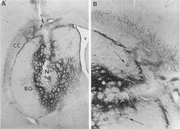
Delivery of adenovirus, herpes simplex virus (HSV), and paramagnetic monocrystalline iron oxide nanoparticles (MION) to rat brain (n = 64) was assessed after intracerebral inoculation or osmotic disruption of the blood-brain barrier (BBB). After intracerebral inoculation, the area of distribution was 7.93 +/- 0.43 mm2 (n = 9) for MION and 9.17 +/- 1.27 mm2 (n = 9) for replication-defective adenovirus. The replication-compromised HSV RH105 spread to 14.00 +/- 0.87 mm2 (n = 8), but also had a large necrotic center (3.54 +/- 0.47 mm2). No infection was detected when virus was administered intra-arterially without hyperosmotic mannitol. After osmotic BBB disruption, delivery of the viruses and MIONs was detected throughout the disrupted cerebral cortex. Positive staining was found in 4 to 845 cells/100 microns thick coronal brain section (n = 7) after adenovirus administration, and in 13 to 197 cells/section (n = 8) after HSV administration. Cells of glial morphology were more frequently stained after administration of adenovirus, whereas neuronal cells were preferentially stained after delivery of both HSV vectors and MION. In a preliminary test of vector delivery in the feline, MION was detected throughout the white matter tracts after inoculation into normal cat brain. Thus MION may be a tool for use in vivo, to monitor the delivery of virus to the central nervous system. Additionally, BBB disruption may be an effective method to globally deliver recombinant viruses to the CNS.
在脑内接种或通过渗透破坏血脑屏障(BBB)后,评估了腺病毒、单纯疱疹病毒(HSV)和顺磁性单晶氧化铁纳米颗粒(MION)向大鼠脑(n = 64)的递送情况。脑内接种后,MION的分布面积为7.93±0.43平方毫米(n = 9),复制缺陷型腺病毒的分布面积为9.17±1.27平方毫米(n = 9)。复制受损的HSV RH105扩散至14.00±0.87平方毫米(n = 8),但也有一个大的坏死中心(3.54±0.47平方毫米)。当在没有高渗甘露醇的情况下动脉内给药病毒时,未检测到感染。在渗透破坏血脑屏障后,在整个被破坏的大脑皮质中检测到病毒和MION的递送。腺病毒给药后,在每100微米厚的冠状脑切片中4至845个细胞中发现阳性染色(n = 7),HSV给药后,在每切片13至197个细胞中发现阳性染色(n = 8)。腺病毒给药后,胶质形态的细胞更频繁地被染色,而在递送HSV载体和MION后,神经元细胞优先被染色。在对猫进行载体递送的初步测试中,将MION接种到正常猫脑后,在整个白质束中都检测到了MION。因此,MION可能是一种用于体内监测病毒向中枢神经系统递送的工具。此外,破坏血脑屏障可能是将重组病毒全局递送至中枢神经系统的有效方法。