Dillon P, Belchis D, Tracy T, Cilley R, Hafer L, Krummel T
Department of Surgery, Pennsylvania State University Children's Hospital, Milton S. Hershey Medical Center, Pennsylvania State University College of Medicine, Hershey.
Am J Pathol. 1994 Aug;145(2):263-7.
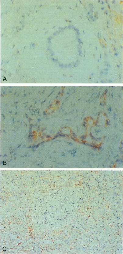
The expression of the inflammatory adhesion molecules intercellular adhesion molecule-1, vascular cell adhesion molecule-1, and endothelial leukocyte adhesion molecule-1, was studied in six infants with biliary atresia using an immunoperoxidase technique on frozen sections. Controls consisted of five patients with various conditions including total parenteral nutrition-induced cholestasis, choledochal cyst, viral hepatitis, metastatic carcinoma, and thrombotic thrombocytopenic purpura. None of the patients were in liver failure. Bile ducts from the control subjects did not express any of the inflammatory adhesion molecules on ductal epithelium. In marked contrast, all of the biliary atresia specimens demonstrated strong intercellular adhesion molecule-1 expression and occasional vascular cell adhesion molecule-1 staining on epithelial cell membranes of both intra- and extrahepatic ductal structures. Hepatocytes and sinusoidal lining cells including Kupffer cells showed a pattern of intense intercellular adhesion molecule-1 and vascular cell adhesion molecule-1 expression in all specimens with active inflammation that could not differentiate the biliary atresia cases from the control group. Lymphocyte function-associated antigen-1 intensely stained the inflammatory cell infiltrate in the biliary atresia and inflamed control specimens. The strong expression of intercellular adhesion molecule-1 on biliary ductal epithelium in patients with biliary atresia suggests a potential role for this adhesion molecule in the pathogenesis of this devastating neonatal hepatic disorder.
采用免疫过氧化物酶技术对6例胆道闭锁婴儿的冰冻切片进行研究,观察炎症黏附分子细胞间黏附分子-1、血管细胞黏附分子-1和内皮细胞白细胞黏附分子-1的表达情况。对照组包括5例患有各种疾病的患者,其中包括全胃肠外营养所致胆汁淤积、胆总管囊肿、病毒性肝炎、转移性癌和血栓性血小板减少性紫癜。所有患者均未出现肝衰竭。对照组患者的胆管在导管上皮上均未表达任何炎症黏附分子。与之形成显著对比的是,所有胆道闭锁标本在肝内和肝外胆管结构的上皮细胞膜上均显示出强烈的细胞间黏附分子-1表达,偶尔还有血管细胞黏附分子-1染色。在所有有活动性炎症的标本中,肝细胞和包括库普弗细胞在内的窦状隙衬里细胞均表现出强烈的细胞间黏附分子-1和血管细胞黏附分子-1表达模式,无法将胆道闭锁病例与对照组区分开来。淋巴细胞功能相关抗原-1在胆道闭锁和炎症对照组标本中的炎性细胞浸润部位呈强染色。胆道闭锁患者胆管上皮细胞间黏附分子-1的强烈表达表明该黏附分子在这种严重的新生儿肝脏疾病发病机制中可能发挥作用。