Pich A, Margaria E, Chiusa L
Department of Biomedical Sciences and Human Oncology, University of Turin, Italy.
Am J Pathol. 1994 Aug;145(2):481-9.
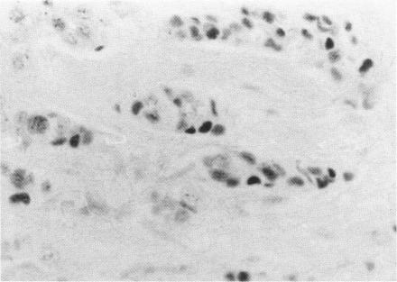
The proliferative activity of male breast carcinoma has been investigated using the staining of the argyrophilic nucleolar organizer regions (AgNORs), the monoclonal antibody against the proliferating cell nuclear antigen (PC10) and the monoclonal antibody MIB-1 in formalin-fixed, paraffin-embedded specimens from 27 primary male breast carcinomas at diagnosis. A significant correlation was found between survival and AgNOR counts (median of survival 77 months for cases with AgNOR/cell < or = 7.27 but 37 months only for cases with > 7.27 AgNOR/cell; P = 0.001), proliferating cell nuclear antigen scores (median of survival 73 months for cases with proliferating cell nuclear antigen < or = 18.25% versus 41 for cases with proliferating cell nuclear antigen > 18.25%; P = 0.013) and MIB-1 scores (median of survival 73 months for cases with MIB-1 scores < or = 23.5% versus 37 months for cases with MIB-1 scores > 23.5%; P = 0.01). Tumor histological grade was also correlated with prognosis (median of survival 72 months for grade 2 versus 33 months for grade 3 tumors; P = 0.01). Estrogen and progesterone receptors, immunohistochemically detected on paraffin-embedded sections, had no prognostic value. In the multivariate survival analysis, only AgNOR counts (P = 0.007) and tumor size (P = 0.003) had an independent prognostic significance. Our results indicate that methods for assessing the cell proliferation in routinely processed specimens offer significant prognostic information in male breast carcinoma. The finding, together with the lack of prognostic significance for estrogen receptors and progesterone receptors, suggests that male breast carcinoma is biologically different from female breast cancer.
利用银染核仁组织区(AgNORs)染色、抗增殖细胞核抗原单克隆抗体(PC10)以及单克隆抗体MIB - 1,对27例原发性男性乳腺癌诊断时福尔马林固定、石蜡包埋标本的增殖活性进行了研究。结果发现,生存率与AgNOR计数(AgNOR/细胞≤7.27的病例中位生存期为77个月,而AgNOR/细胞>7.27的病例仅为37个月;P = 0.001)、增殖细胞核抗原评分(增殖细胞核抗原≤18.25%的病例中位生存期为73个月,而增殖细胞核抗原>18.25%的病例为41个月;P = 0.013)以及MIB - 1评分(MIB - 1评分≤23.5%的病例中位生存期为73个月,而MIB - 1评分>23.5%的病例为37个月;P = 0.01)之间存在显著相关性。肿瘤组织学分级也与预后相关(2级肿瘤中位生存期为72个月,3级肿瘤为33个月;P = 0.01)。在石蜡包埋切片上通过免疫组织化学检测的雌激素和孕激素受体无预后价值。在多因素生存分析中,只有AgNOR计数(P = 0.007)和肿瘤大小(P = 0.003)具有独立的预后意义。我们的结果表明,评估常规处理标本中细胞增殖的方法可为男性乳腺癌提供重要的预后信息。这一发现,连同雌激素受体和孕激素受体缺乏预后意义,提示男性乳腺癌在生物学上与女性乳腺癌不同。