Rodgers D W, Gamblin S J, Harris B A, Ray S, Culp J S, Hellmig B, Woolf D J, Debouck C, Harrison S C
Department of Molecular and Cellular Biology, Harvard University, Cambridge, MA 02138.
Proc Natl Acad Sci U S A. 1995 Feb 14;92(4):1222-6. doi: 10.1073/pnas.92.4.1222.
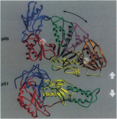
The crystal structure of the reverse transcriptase (RT) from the type 1 human immunodeficiency virus has been determined at 3.2-A resolution. Comparison with complexes between RT and the polymerase inhibitor Nevirapine [Kohlstaedt, L.A., Wang, J., Friedman, J.M., Rice, P.A. & Steitz, T.A. (1992) Science 256, 1783-1790] and between RT and an oligonucleotide [Jacobo-Molina, A., Ding, J., Nanni, R., Clark, A. D., Lu, X., Tantillo, C., Williams, R. L., Kamer, G., Ferris, A. L., Clark, P., Hizi, A., Hughes, S. H. & Arnold, E. (1993) Proc. Natl. Acad. Sci. USA 90, 6320-6324] reveals changes associated with ligand binding. The enzyme is a heterodimer (p66/p51), with domains labeled "fingers," "thumb," "palm," and "connection" in both subunits, and a ribonuclease H domain in the larger subunit only. The most striking difference between RT and both complex structures is the change in orientation of the p66 thumb (approximately 33 degrees rotation). Smaller shifts relative to the core of the molecule were also found in other domains, including the p66 fingers and palm, which contain the polymerase active site. Within the polymerase catalytic region itself, there are no rearrangements between RT and the RT/DNA complex. In RT/Nevirapine, the drug binds in the p66 palm near the polymerase active site, a region that is well-packed hydrophobic core in the unliganded enzyme. Room for the drug is provided by movement of a small beta-sheet within the palm domain of the Nevirapine complex. The rearrangement within the palm and thumb, as well as domain shifts relative to the enzyme core, may prevent correct placement of the oligonucleotide substrate when the drug is bound.
已确定1型人类免疫缺陷病毒逆转录酶(RT)的晶体结构,分辨率为3.2埃。将其与RT和聚合酶抑制剂奈韦拉平之间的复合物[科尔施泰特,L.A.,王,J.,弗里德曼,J.M.,赖斯,P.A. & 施泰茨,T.A.(1992年)《科学》256卷,1783 - 1790页]以及RT和寡核苷酸之间的复合物[雅各布 - 莫利纳,A.,丁,J.,南尼,R.,克拉克,A.D.,卢,X.,坦蒂洛,C.,威廉姆斯,R.L.,卡默,G.,费里斯,A.L.,克拉克,P.,希齐,A.,休斯,S.H. & 阿诺德,E.(1993年)《美国国家科学院院刊》90卷,6320 - 6324页]进行比较,揭示了与配体结合相关的变化。该酶是一种异二聚体(p66/p51),两个亚基中均有标记为“手指”“拇指”“手掌”和“连接”的结构域,且仅在较大亚基中有一个核糖核酸酶H结构域。RT与两种复合物结构之间最显著的差异是p66拇指的方向变化(约33度旋转)。在包括含有聚合酶活性位点的p66手指和手掌在内的其他结构域中,相对于分子核心也发现了较小的位移。在聚合酶催化区域本身,RT与RT/DNA复合物之间没有重排。在RT/奈韦拉平复合物中,药物结合在靠近聚合酶活性位点的p66手掌中,该区域在未结合配体的酶中是紧密堆积的疏水核心。奈韦拉平复合物手掌结构域内一个小β折叠的移动为药物提供了空间。手掌和拇指内的重排以及相对于酶核心的结构域位移,可能会在药物结合时阻止寡核苷酸底物的正确定位。