Wu C C, Croxtall J D, Perretti M, Bryant C E, Thiemermann C, Flower R J, Vane J R
Department of Vascular Biology, William Harvey Research Institute, St. Bartholomew's Hospital Medical College, London, United Kingdom.
Proc Natl Acad Sci U S A. 1995 Apr 11;92(8):3473-7. doi: 10.1073/pnas.92.8.3473.
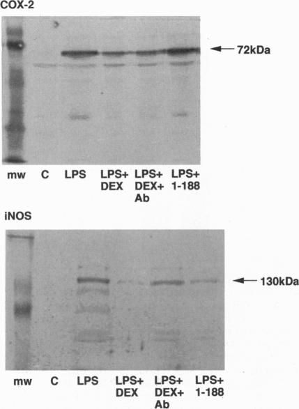
Administration of Escherichia coli lipopolysaccharide (LPS; 10 mg/kg i.v.) to male Wistar rats caused within 240 min (i) a sustained fall (approximately 30 mmHg) in mean arterial blood pressure, (ii) a reduction (> 75%) in the pressor responses to norepinephrine (1 microgram/kg i.v.), and (iii) an induction of nitric oxide synthase (iNOS) as measured in the lung. Dexamethasone (1 mg/kg i.p. at 2 h prior to LPS) attenuated the hypotension and the vascular hyporeactivity to norepinephrine and reduced (by approximately 77%) the expression of iNOS in the lung. These effects of dexamethasone were prevented by pretreatment of LPS-treated rats with a neutralizing antiserum to lipocortin 1 (anti-LC1; 60 mg/kg s.c. at 24 h prior to LPS) but not by a control nonimmune sheep serum. Stimulation of J774.2 macrophages with LPS (1 microgram/ml for 24 h) caused the expression of iNOS and cyclooxygenase 2 (COX-2) protein and significantly increased nitrite generation; this was prevented by dexamethasone (0.1 microM at 1 h prior to LPS), which also increased cell surface lipocortin 1. Pretreatment of J774.2 cells with anti-LC1 (1:60 dilution at 4 h prior to LPS) also abolished the inhibitory effect of dexamethasone on iNOS expression and nitrite accumulation but not that on COX-2 expression. A lipocortin 1 fragment (residues 1-188 of human lipocortin 1; 20 micrograms/ml at 1 h prior to LPS) also blocked iNOS in J774.2 macrophages activated by LPS (approximately 78% inhibition), and this too was prevented by anti-LC1. We conclude that the extracellular release of endogenous lipocortin 1 (i) mediates the inhibition by dexamethasone of the expression of iNOS, but not of COX-2, and (ii) contributes substantially to the beneficial actions of dexamethasone in rats with endotoxic shock.
给雄性Wistar大鼠静脉注射大肠杆菌脂多糖(LPS;10mg/kg),在240分钟内导致:(i)平均动脉血压持续下降(约30mmHg);(ii)对去甲肾上腺素(1μg/kg静脉注射)的升压反应降低(>75%);(iii)肺中一氧化氮合酶(iNOS)的诱导。地塞米松(在LPS注射前2小时腹腔注射1mg/kg)减轻了低血压和对去甲肾上腺素的血管低反应性,并降低了肺中iNOS的表达(约77%)。用抗脂皮质素1中和抗血清(LPS注射前24小时皮下注射60mg/kg)预处理LPS处理的大鼠可防止地塞米松的这些作用,但对照非免疫绵羊血清则不能。用LPS(1μg/ml处理24小时)刺激J774.2巨噬细胞可导致iNOS和环氧化酶2(COX-2)蛋白的表达,并显著增加亚硝酸盐的生成;这可被地塞米松(LPS注射前1小时0.1μM)阻止,地塞米松还增加了细胞表面脂皮质素1。用抗LC1(LPS注射前4小时1:60稀释)预处理J774.2细胞也消除了地塞米松对iNOS表达和亚硝酸盐积累的抑制作用,但对COX-2表达的抑制作用未消除。脂皮质素1片段(人脂皮质素1的1-188位残基;LPS注射前1小时20μg/ml)也可阻断LPS激活的J774.2巨噬细胞中的iNOS(约78%抑制),这也可被抗LC1阻止。我们得出结论,内源性脂皮质素1的细胞外释放:(i)介导地塞米松对iNOS表达的抑制,但不介导对COX-2表达的抑制;(ii)在很大程度上有助于地塞米松对内毒素休克大鼠的有益作用。