Singer I I, Kawka D W, Bayne E K, Donatelli S A, Weidner J R, Williams H R, Ayala J M, Mumford R A, Lark M W, Glant T T
Division of Immunology and Inflammation, Merck Research Laboratories, Merck & Co., Inc., Rahway, New Jersey 07065, USA.
J Clin Invest. 1995 May;95(5):2178-86. doi: 10.1172/JCI117907.
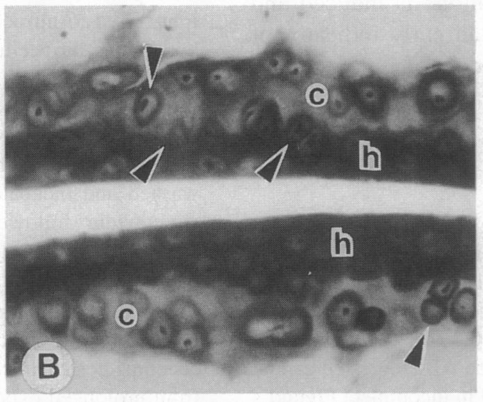
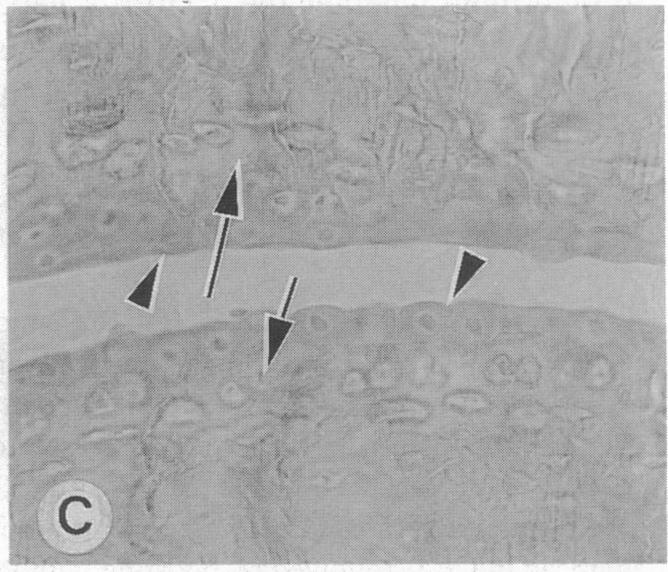
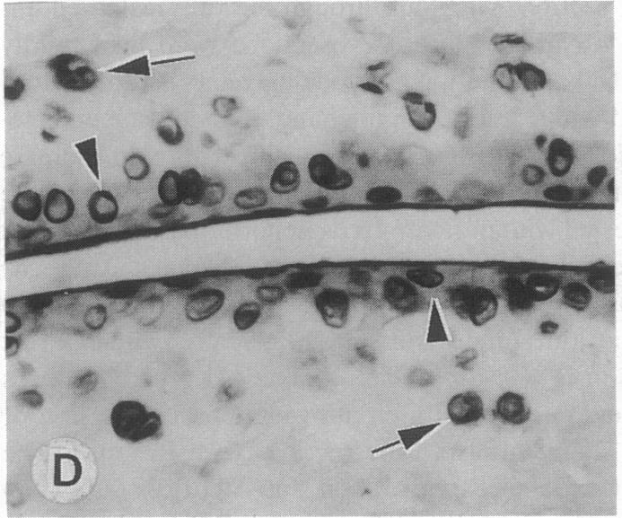
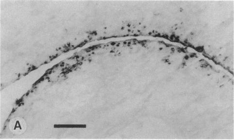
The destruction of articular cartilage in immune inflammatory arthritic disease involves the proteolytic degradation of its extracellular matrix. The role of activated matrix metalloproteinases (MMPs) in the chondrodestructive process was studied by identifying a selective cleavage product of aggrecan in murine arthritis models initiated by immunization with either type II collagen or proteoglycan. We conducted semiquantitative immunocytochemical studies of VDIPEN341 using a monospecific polyclonal antibody requiring the free COOH group of the COOH-terminal Asn for epitope detection. This antibody recognizes the aggrecan G1 domain fragment generated by MMP [i.e., stromelysin (SLN) or gelatinase A] cleavage of aggrecan between Asn341-Phe342 but does not recognize intact aggrecan. VDIPEN was undetectable in normal mouse cartilage but was observed in the articular cartilage (AC) of mice with collagen-induced arthritis 10 d after immunization, without histological damage and clinical symptoms. This aggrecan neoepitope was colocalized with high levels of glycosaminoglycans (GAGs) in pericellular matrices of AC chondrocytes but was not seen at the articular surface at this early time. Digestion of normal (VDIPEN negative) mouse paw cryosections with SLN also produced heavy pericellular VDIPEN labeling. Computer-based image analysis showed that the amount of VDIPEN expression increased dramatically by 20 d (70% of the SLN maximum) and was correlated with GAG depletion. Both infiltration of inflammatory cells into the synovial cavity and early AC erosion were also very prominent at this time. Analysis of adjacent sections showed that both induction of VDIPEN and GAG depletion were strikingly codistributed within sites of articular cartilage damage. Similar results occurred in proteoglycan-induced arthritis, a more progressive and chronic model of inflammatory arthritis. These studies demonstrate for the first time the MMP-dependent catabolism of aggrecan at sites of chondrodestruction during inflammatory arthritis.
免疫炎性关节疾病中关节软骨的破坏涉及细胞外基质的蛋白水解降解。通过在由II型胶原蛋白或蛋白聚糖免疫引发的小鼠关节炎模型中鉴定聚集蛋白聚糖的选择性裂解产物,研究了活化的基质金属蛋白酶(MMPs)在软骨破坏过程中的作用。我们使用一种单特异性多克隆抗体对VDIPEN341进行了半定量免疫细胞化学研究,该抗体需要COOH末端Asn的游离COOH基团来检测表位。该抗体识别由MMP [即基质溶解素(SLN)或明胶酶A]在Asn341 - Phe342之间裂解聚集蛋白聚糖产生的聚集蛋白聚糖G1结构域片段,但不识别完整的聚集蛋白聚糖。VDIPEN在正常小鼠软骨中无法检测到,但在免疫后10天的胶原诱导性关节炎小鼠的关节软骨(AC)中观察到,此时没有组织学损伤和临床症状。这种聚集蛋白聚糖新表位与AC软骨细胞周围基质中高水平的糖胺聚糖(GAGs)共定位,但在这个早期阶段在关节表面未观察到。用SLN消化正常(VDIPEN阴性)小鼠爪冷冻切片也产生了大量的细胞周围VDIPEN标记。基于计算机的图像分析表明,VDIPEN表达量在20天时急剧增加(达到SLN最大值的70%),并且与GAG消耗相关。此时,炎性细胞向滑膜腔的浸润和早期AC侵蚀也非常明显。相邻切片分析表明,VDIPEN的诱导和GAG消耗在关节软骨损伤部位显著共分布。在蛋白聚糖诱导的关节炎中也出现了类似结果,这是一种更进展性和慢性的炎性关节炎模型。这些研究首次证明了在炎性关节炎期间软骨破坏部位聚集蛋白聚糖的MMP依赖性分解代谢。