deSouza N M, Kmiot W A, Puni R, Hall A S, Burl M, Bartram C I, Bydder G M
Robert Steiner Magnetic Resonance Unit, Department of Radiology, Royal Postgraduate Medical School, London.
Gut. 1995 Aug;37(2):284-7. doi: 10.1136/gut.37.2.284.
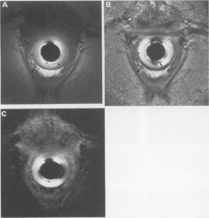
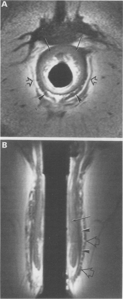
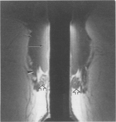
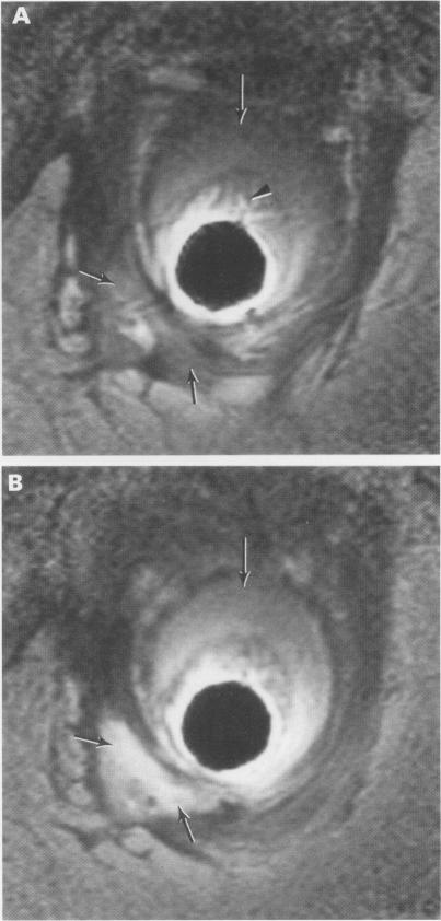
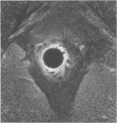
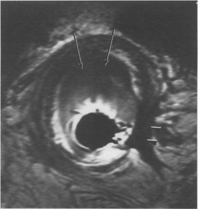
An internal receiver coil was used to obtain high resolution transverse and oblique coronal magnetic resonance images of the anal sphincter in five normal volunteers and five patients. The internal sphincter had a high signal intensity on T1 weighted, T2 weighted, and STIR sequences whereas the conjoined longitudinal muscle and external sphincter had a low signal intensity. The internal sphincter (but not the external sphincter) showed contrast enhancement after administration of intravenous gadopentetate dimeglumine. The oblique coronal plane was particularly useful for showing the thickness and the relations of the external sphincter. Sphincteric abscesses as well as muscle defects, hypertrophy, and atrophy were clearly shown. The coil was well tolerated by most subjects. It has considerable potential for improving the diagnosis of anorectal disease.
使用内置接收线圈对5名正常志愿者和5名患者的肛门括约肌进行高分辨率横向和斜冠状面磁共振成像。内括约肌在T1加权、T2加权和短TI反转恢复(STIR)序列上呈高信号强度,而联合纵肌和外括约肌呈低信号强度。静脉注射钆喷酸葡胺后,内括约肌(而非外括约肌)显示出对比增强。斜冠状面对于显示外括约肌的厚度及其关系特别有用。括约肌脓肿以及肌肉缺损、肥大和萎缩均清晰可见。大多数受试者对该线圈耐受性良好。它在改善肛肠疾病的诊断方面具有相当大的潜力。