Stolinski C
Department of Anatomy and Cell Biology, St Mary's Hospital Medical School, Imperial College of Science, Technology and Medicine, London, UK.
J Anat. 1995 Jun;186 ( Pt 3)(Pt 3):577-83.
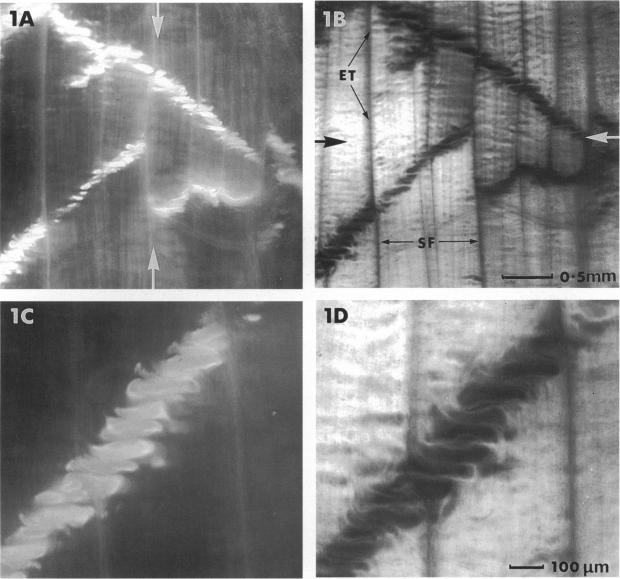
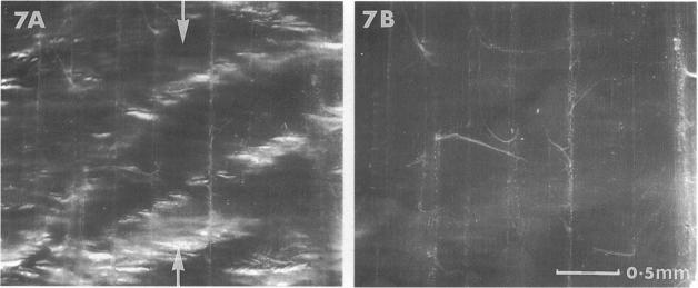
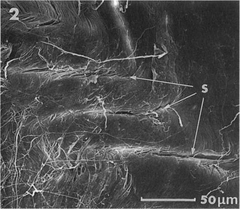
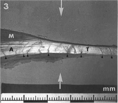
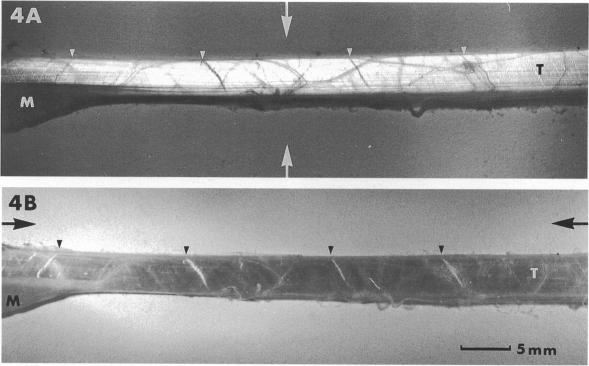
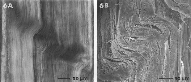
Fixed and unfixed human tendons originating from cadavers and postoperation specimens were examined using inclined parallel beams of light in a reflecting mode. Along the tendon, numerous planes, constantly inclined to the axis, were observed edge-on at the surface and within the interior. Their angle of inclination, with respect to the distal end was very nearly +/- 50 degrees. The planes consisted of individual segments arranged in steps which were on average 190 x 50 microns. Similar configurations were also observed with the scanning electron microscope. Using this technique, the segments were identified with collagen bundles turning at a sharp angle with respect to the axis of the tendon at the level of the inclined plane. Crimped planes were found to be irregularly distributed along the tendons. On longer flatter tendons the average distance between planes was in the range of 1-12 mm. On stretching, the inclined pattern disappeared and was rapidly reestablished in the previously observed position when the strain was released. It is suggested that the observed structure forms a mechanism which is responsible for the appearance of the first part of 'foot' region of the tendon's stress-strain diagram.
使用反射模式下的倾斜平行光束对来自尸体和术后标本的固定和未固定人体肌腱进行了检查。沿着肌腱,在表面和内部都能观察到许多与轴线呈恒定倾斜的平面的边缘。它们相对于远端的倾斜角度非常接近±50度。这些平面由平均为190×50微米的呈阶梯状排列的单个节段组成。用扫描电子显微镜也观察到了类似的结构。利用这项技术,这些节段被确定为在倾斜平面处胶原束相对于肌腱轴线急剧转折。发现卷曲平面沿肌腱不规则分布。在较长较平的肌腱上,平面之间的平均距离在1 - 12毫米范围内。拉伸时,倾斜模式消失,当应变释放时又迅速在先前观察到的位置重新建立。有人认为,观察到的结构形成了一种机制,该机制负责肌腱应力 - 应变图“足部”区域第一部分的出现。