Taylor K A, Taylor D W
Department of Cell Biology, Duke University Medical Center, Durham, North Carolina 27710.
Biophys J. 1994 Nov;67(5):1976-83. doi: 10.1016/S0006-3495(94)80680-0.
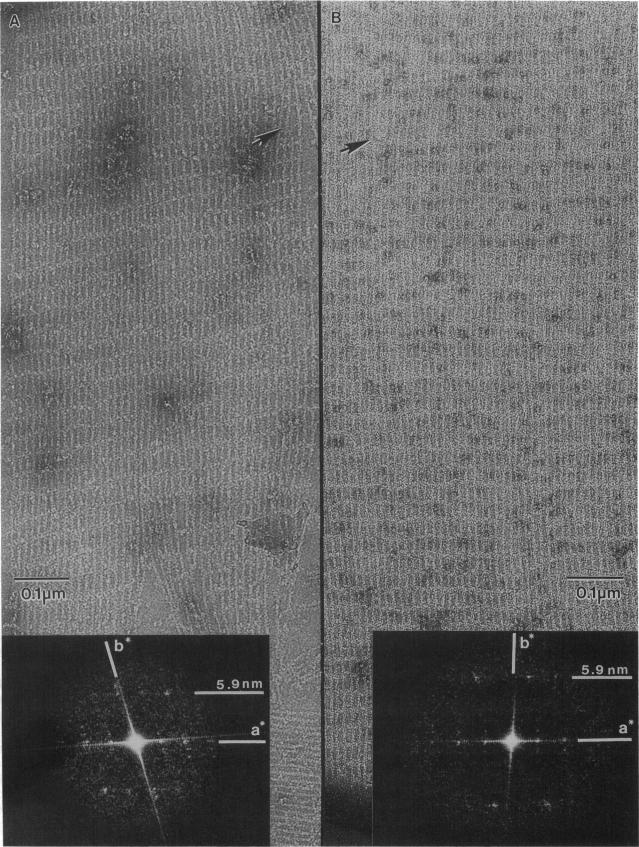
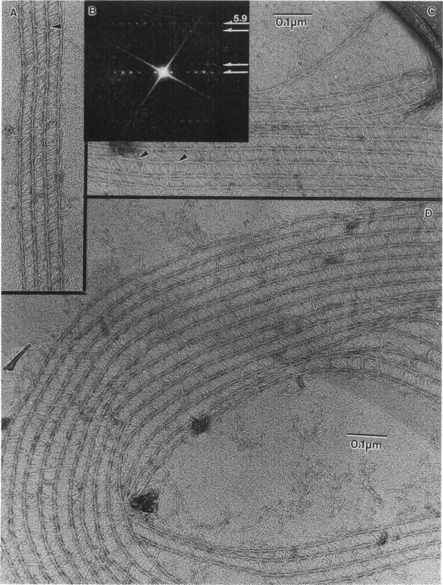
A method is described for forming two-dimensional (2-D) paracrystalline complexes of F-actin and bundling/gelation proteins on positively charged lipid monolayers. These arrays facilitate detailed structural studies of protein interactions with F-actin by eliminating superposition effects present in 3-D bundles. Bundles of F-actin have been produced using the glycolytic enzymes aldolase and glyceraldehyde-3-phosphate dehydrogenase, the cytoskeletal protein erythrocyte adducin as well as smooth muscle alpha-actinin from chicken gizzard. All of the 2-D bundles formed contain F-actin with a 13/6 helical structure. F-actin-aldolase bundles have an interfilament spacing of 12.6 nm and a superlattice arrangement of actin filaments that can be explained by expression of a local twofold axis in the neighborhood of the aldolase. Well ordered F-actin-alpha-actinin 2-D bundles have an interfilament spacing of 36 nm and contain crosslinks 33 nm in length angled approximately 25-35 degrees to the filament axis. Images and optical diffraction patterns of these bundles suggest that they consist of parallel, unipolar arrays of actin filaments. This observation is consistent with an actin crosslinking function at adhesion plaques where actin filaments are bound to the cell membrane with uniform polarity.
本文描述了一种在带正电荷的脂质单层上形成F-肌动蛋白与成束/凝胶化蛋白的二维(2-D)准晶复合物的方法。这些阵列通过消除三维束中存在的叠加效应,有助于对蛋白质与F-肌动蛋白相互作用进行详细的结构研究。已使用糖酵解酶醛缩酶和甘油醛-3-磷酸脱氢酶、细胞骨架蛋白红细胞内收蛋白以及来自鸡砂囊的平滑肌α-辅肌动蛋白产生了F-肌动蛋白束。形成的所有二维束均含有具有13/6螺旋结构的F-肌动蛋白。F-肌动蛋白-醛缩酶束的丝间间距为12.6 nm,肌动蛋白丝的超晶格排列可以通过醛缩酶附近局部二重轴的表达来解释。排列良好的F-肌动蛋白-α-辅肌动蛋白二维束的丝间间距为36 nm,含有长度为33 nm的交联,与丝轴成约25-35度角。这些束的图像和光学衍射图表明它们由平行、单极的肌动蛋白丝阵列组成。这一观察结果与肌动蛋白在黏着斑处的交联功能一致,在黏着斑处肌动蛋白丝以均匀的极性与细胞膜结合。