Li G, Rungger-Brändle E, Just I, Jonas J C, Aktories K, Wollheim C B
Department of Medicine, University of Geneva, Switzerland.
Mol Biol Cell. 1994 Nov;5(11):1199-213. doi: 10.1091/mbc.5.11.1199.
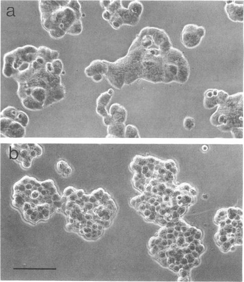
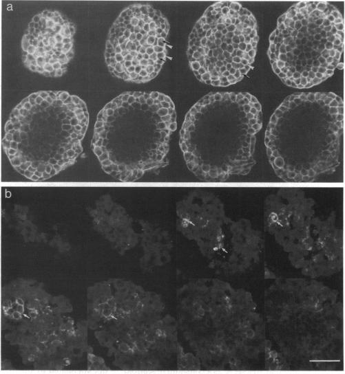
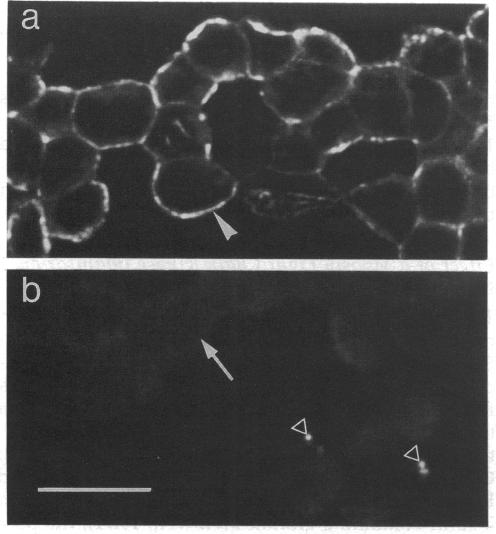
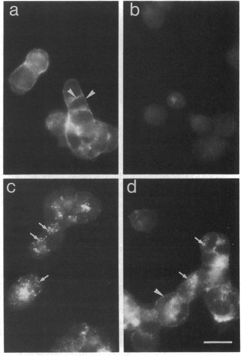
To examine their role in insulin secretion, actin filaments (AFs) were disrupted by Clostridium botulinum C2 toxin that ADP-ribosylates G-actin. Ribosylation also prevents polymerization of G-actin to F-actin and inhibits AF assembly by capping the fast-growing end of F-actin. Pretreatment of HIT-T15 cells with the toxin inhibited stimulated insulin secretion in a time- and dose-dependent manner. The toxin did not affect cellular insulin content or nonstimulated secretion. In static incubation, toxin treatment caused 45-50% inhibition of secretion induced by nutrients alone (10 mM glucose + 5 mM glutamine + 5 mM leucine) or combined with bombesin (phospholipase C-activator) and 20% reduction of that potentiated by forskolin (stimulator of adenylyl cyclase). In perifusion, the stimulated secretion during the first phase was marginally diminished, whereas the second phase was inhibited by approximately 80%. Pretreatment of HIT cells with wartmannin, a myosin light chain kinase inhibitor, caused a similar pattern of inhibition of the biphasic insulin release as C2 toxin. Nutrient metabolism and bombesin-evoked rise in cytosolic free Ca2+ were not affected by C2 toxin, indicating that nutrient recognition and the coupling between receptor activation and second messenger generation was not changed. In the toxin-treated cells, the AF web beneath the plasma membrane and the diffuse cytoplasmic F-actin fibers disappeared, as shown both by staining with an antibody against G- and F-actin and by staining F-actin with fluorescent phallacidin. C2 toxin dose-dependently reduced cellular F-actin content. Stimulation of insulin secretion was not associated with changes in F-actin content and organization. Treatment of cells with cytochalasin E and B, which shorten AFs, inhibited the stimulated insulin release by 30-50% although differing in their effects on F-actin content. In contrast to HIT-T15 cells, insulin secretion was potentiated in isolated rat islets after disruption of microfilaments with C2 toxin, most notably during the first phase. This effect was, however, diminished, and the second phase became slightly inhibited when the islets were degranulated. These results indicate an important role for AFs in insulin secretion. In the poorly granulated HIT-T15 cells actin-myosin interactions may participate in the recruitment of secretory granules to the releasable pool. In native islet beta-cells the predominant function of AFs appears to be the limitation of the access of granules to the plasma membrane.
为了研究肌动蛋白丝(AFs)在胰岛素分泌中的作用,肉毒杆菌C2毒素破坏了AFs,该毒素可使G-肌动蛋白进行ADP核糖基化。核糖基化还可阻止G-肌动蛋白聚合成F-肌动蛋白,并通过封闭F-肌动蛋白快速生长的末端来抑制AF组装。用该毒素预处理HIT-T15细胞,可呈时间和剂量依赖性地抑制刺激后的胰岛素分泌。该毒素不影响细胞内胰岛素含量或基础分泌。在静态孵育中,毒素处理使单独由营养物质(10 mM葡萄糖 + 5 mM谷氨酰胺 + 5 mM亮氨酸)诱导的分泌或与蛙皮素(磷脂酶C激活剂)联合诱导的分泌受到45 - 50%的抑制,使由福斯可林(腺苷酸环化酶刺激剂)增强的分泌减少20%。在灌流实验中,第一相的刺激分泌略有减少,而第二相则受到约80%的抑制。用肌球蛋白轻链激酶抑制剂渥曼青霉素预处理HIT细胞,导致双相胰岛素释放的抑制模式与C2毒素相似。营养物质代谢以及蛙皮素引起的胞质游离Ca2+升高不受C2毒素影响,这表明营养物质识别以及受体激活与第二信使产生之间的偶联未发生改变。在用毒素处理的细胞中,质膜下方的AF网络以及弥漫性的胞质F-肌动蛋白纤维消失,这通过抗G-和F-肌动蛋白抗体染色以及用荧光鬼笔环肽对F-肌动蛋白染色均得以显示。C2毒素呈剂量依赖性地降低细胞内F-肌动蛋白含量。胰岛素分泌的刺激与F-肌动蛋白含量和组织的变化无关。用细胞松弛素E和B处理细胞,它们可缩短AFs,尽管对F-肌动蛋白含量的影响不同,但可使刺激后的胰岛素释放受到30 - 50%的抑制。与HIT-T15细胞不同,用C2毒素破坏微丝后,分离的大鼠胰岛中的胰岛素分泌增强,最显著的是在第一相。然而,当胰岛脱颗粒时,这种作用减弱,第二相略有抑制。这些结果表明AFs在胰岛素分泌中起重要作用。在颗粒较少的HIT-T15细胞中,肌动蛋白-肌球蛋白相互作用可能参与分泌颗粒募集到可释放池的过程。在天然胰岛β细胞中,AFs的主要功能似乎是限制颗粒接近质膜。