Zaki S R, Greer P W, Coffield L M, Goldsmith C S, Nolte K B, Foucar K, Feddersen R M, Zumwalt R E, Miller G L, Khan A S
Division of Viral and Rickettsial Diseases, Centers for Disease Control and Prevention, Atlanta, Georgia 30333.
Am J Pathol. 1995 Mar;146(3):552-79.
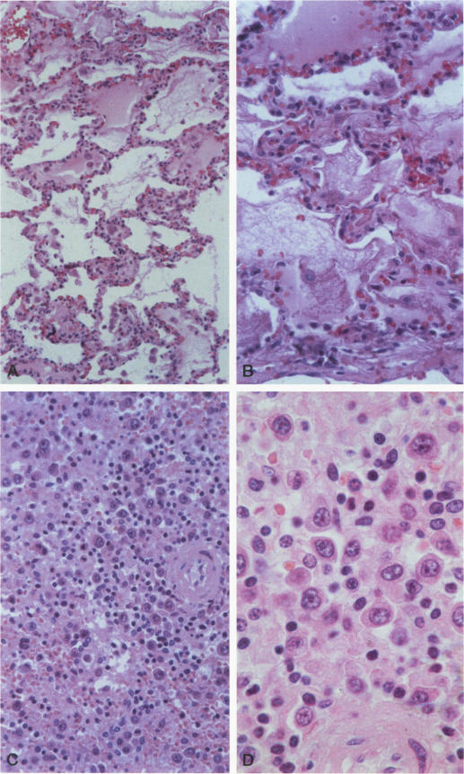
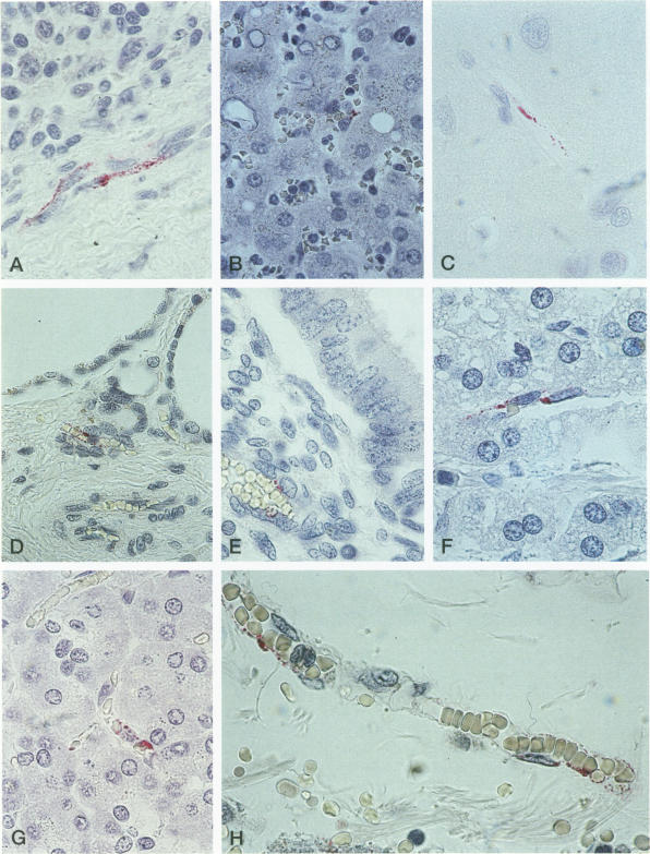
A recent outbreak of a severe pulmonary disease in the southwestern United States was etiologically linked to a previously unrecognized hantavirus. The virus has been isolated from its major reservoir, the deer mouse, Peromyscus maniculatus, and recently named Sin Nombre virus. Clinically, the disease has become known as the hantavirus pulmonary syndrome (HPS). Since May 1993, 44 fatal cases of HPS have been identified through clinicopathological review and immunohistochemical (IHC) testing of tissues from 273 patients who died of an unexplained noncardiogenic pulmonary edema. In 158 cases for which suitable specimens were available, serological testing and/or reverse transcription-polymerase chain reaction (RT-PCR) amplification of extracted RNA was also performed. IHC, serological, and PCR results were concordant for virtually all HPS and non-HPS patients when more than one assay was performed. The prodromal illness of HPS is similar to that of many other viral diseases. Consistent hematological features include thrombocytopenia, hemoconcentration, neutrophilic leukocytosis with a left shift, and reactive lymphocytes. Pulmonary histopathological features were similar in most of the fatal HPS cases (40/44) and consisted of an interstitial pneumonitis with a variable mononuclear cell infiltrate, edema, and focal hyaline membranes. In four cases, however, pulmonary features were significantly different and included diffuse alveolar damage and variable degrees of severe air space disorganization. IHC analysis showed widespread presence of hantaviral antigens in endothelial cells of the microvasculature, particularly in the lung. Hantaviral antigens were also observed within follicular dendritic cells, macrophages, and lymphocytes. Hantaviral inclusions were observed in endothelial cells of lungs by thinsection electron microscopy, and their identity was verified by immunogold labeling. Virus-like particles were seen in pulmonary endothelial cells and macrophages. HPS is a newly recognized, often fatal disease, with a spectrum of microscopic morphological changes, which may be an important cause of severe and fatal illness presenting as adult respiratory distress syndrome.
美国西南部最近爆发的一种严重肺部疾病在病因上与一种此前未被识别的汉坦病毒有关。该病毒已从其主要宿主鹿鼠(白足鼠)中分离出来,并于近期被命名为辛诺柏病毒。临床上,这种疾病已被称为汉坦病毒肺综合征(HPS)。自1993年5月以来,通过对273例死于不明原因非心源性肺水肿患者的组织进行临床病理检查和免疫组织化学(IHC)检测,已确诊44例HPS死亡病例。对于158例有合适标本的病例,还进行了血清学检测和/或对提取RNA的逆转录聚合酶链反应(RT-PCR)扩增。当进行不止一种检测时,几乎所有HPS患者和非HPS患者的IHC、血清学和PCR结果都是一致的。HPS的前驱疾病与许多其他病毒疾病相似。一致的血液学特征包括血小板减少、血液浓缩、中性粒细胞增多伴核左移以及反应性淋巴细胞。大多数致命HPS病例(40/44)的肺部组织病理学特征相似,表现为间质性肺炎,伴有不同程度的单核细胞浸润、水肿和局灶性透明膜形成。然而,有4例患者的肺部特征明显不同,包括弥漫性肺泡损伤和不同程度的严重气腔结构紊乱。IHC分析显示,汉坦病毒抗原广泛存在于微血管的内皮细胞中,尤其是在肺部。在滤泡树突状细胞、巨噬细胞和淋巴细胞内也观察到汉坦病毒抗原。通过超薄切片电子显微镜在肺内皮细胞中观察到汉坦病毒包涵体,并通过免疫金标记验证了其身份。在肺内皮细胞和巨噬细胞中可见病毒样颗粒。HPS是一种新发现的、通常致命的疾病,具有一系列微观形态学变化,可能是导致表现为成人呼吸窘迫综合征的严重致命疾病的重要原因。