Yoshida K, Kaji M, Takahashi T, van den Berg T K, Dijkstra C D
Department of Pathology, Tohoku University, Sendai, Japan.
Immunology. 1995 Jan;84(1):117-26.
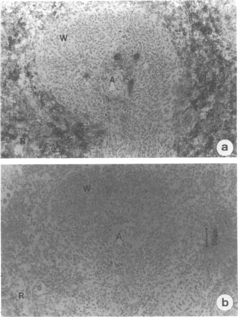
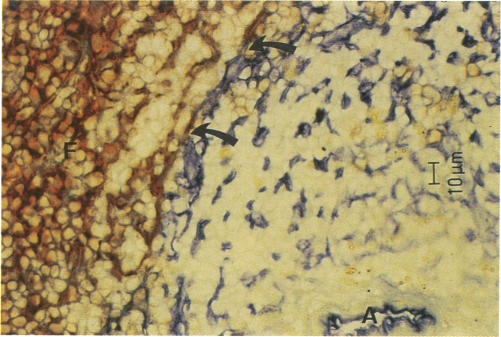
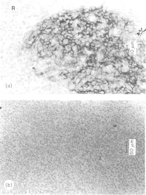
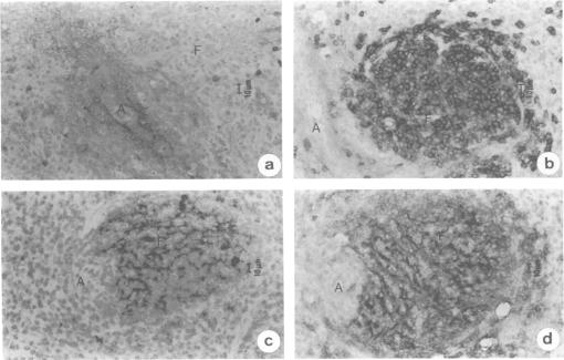
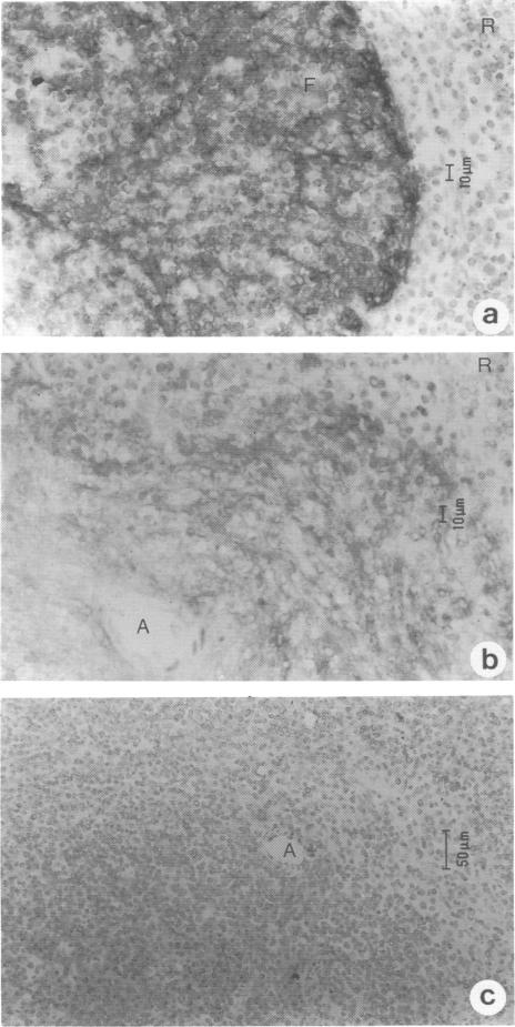
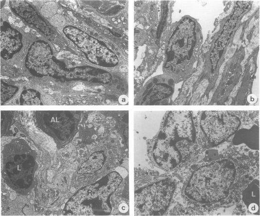
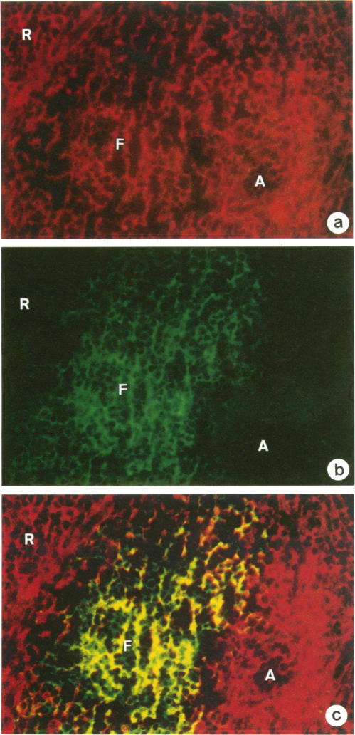
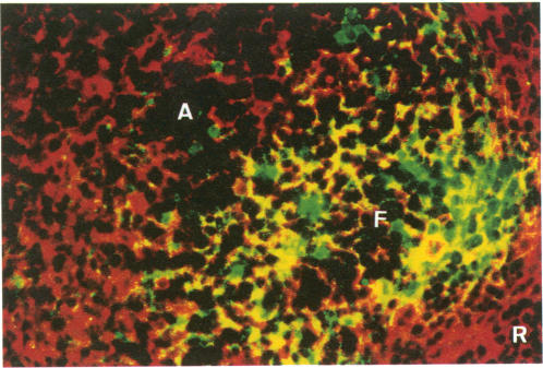
Follicular dendritic cells (FDC) are uniquely characterized by the ability to trap immune complexes. In a previous report, it was shown that functional FDC with the capacity to trap immune complexes via complement receptor emerged in the splenic follicle after transferring syngeneic lymphocytes into the severe combined immunodeficiency (SCID) mouse. In the present report, we have investigated whether FDC are derived from haematopoietic cells or surrounding stromal components, by transferring allogeneic lymphocytes into SCID mice. Transfer of allogeneic T and B lymphocytes (H-2k) into SCID(H-2d) mice, however, failed to induce the development of FDC in the splenic white pulp. This was due to a graft-versus-host reaction (GVHR) by allogeneic lymphocytes against host stromal cells, as revealed by the destruction of the splenic reticular meshwork. The GVHR was prevented in transfer experiments of T-cell-depleted allogeneic lymphocytes with daily administration of anti-Thy-1 antibody. This resulted in segregated lodgement of allogeneic B lymphocytes in the proper compartments and, thereafter, generation of FDC in the primary follicle of SCID spleen, as revealed by the trapped immune complexes via complement receptors. The H-2 of the newly generated FDC was examined by two-colour immunofluorescent staining. FDC were defined as the reticular cells stained with anti-CR1/2 or FDC-M1 antibodies. FDC carried host H-2, clearly indicating that newly generated FDC are host-derived. In addition, the FDC shared the BP-3 protein with the surrounding reticular cells, a specific marker of reticular meshwork in the murine lymphoid tissues, and formed a network continuous with the rest of the reticulum, suggesting that FDC and non-FDC reticular cells belong to the same cell lineage.
滤泡树突状细胞(FDC)的独特特征是能够捕获免疫复合物。在先前的一份报告中显示,将同基因淋巴细胞转移到严重联合免疫缺陷(SCID)小鼠体内后,脾脏滤泡中出现了具有通过补体受体捕获免疫复合物能力的功能性FDC。在本报告中,我们通过将同种异体淋巴细胞转移到SCID小鼠体内,研究了FDC是源自造血细胞还是周围的基质成分。然而,将同种异体T和B淋巴细胞(H-2k)转移到SCID(H-2d)小鼠体内未能诱导脾脏白髓中FDC的发育。这是由于同种异体淋巴细胞对宿主基质细胞的移植物抗宿主反应(GVHR),脾脏网状结构的破坏表明了这一点。在T细胞耗竭的同种异体淋巴细胞转移实验中,通过每日给予抗Thy-1抗体可预防GVHR。这导致同种异体B淋巴细胞在适当的隔室中分离定位,此后,SCID脾脏初级滤泡中产生FDC,通过补体受体捕获的免疫复合物可证明这一点。通过双色免疫荧光染色检查新生成的FDC的H-2。FDC被定义为用抗CR1/2或FDC-M1抗体染色的网状细胞。FDC携带宿主H-2,清楚地表明新生成的FDC源自宿主。此外,FDC与周围的网状细胞共享BP-3蛋白,BP-3蛋白是小鼠淋巴组织中网状结构的特异性标志物,并且与其余的网状结构形成连续的网络,这表明FDC和非FDC网状细胞属于同一细胞谱系。