• 文献检索
  • 文档翻译
  • 深度研究
  • 学术资讯
  • Suppr Zotero 插件Zotero 插件
  • 邀请有礼
  • 套餐&价格
  • 历史记录
应用&插件
Suppr Zotero 插件Zotero 插件浏览器插件Mac 客户端Windows 客户端微信小程序
定价
高级版会员购买积分包购买API积分包
服务
文献检索文档翻译深度研究API 文档MCP 服务
关于我们
关于 Suppr公司介绍联系我们用户协议隐私条款
关注我们

Suppr 超能文献

核心技术专利:CN118964589B侵权必究
粤ICP备2023148730 号-1Suppr @ 2026

文献检索

告别复杂PubMed语法,用中文像聊天一样搜索,搜遍4000万医学文献。AI智能推荐,让科研检索更轻松。

立即免费搜索

文件翻译

保留排版,准确专业,支持PDF/Word/PPT等文件格式,支持 12+语言互译。

免费翻译文档

深度研究

AI帮你快速写综述,25分钟生成高质量综述,智能提取关键信息,辅助科研写作。

立即免费体验

三种细胞周期相关抗原作为乳腺癌增殖活性和预后标志物的比较

Comparison of three cell cycle associated antigens as markers of proliferative activity and prognosis in breast carcinoma.

作者信息

Gillett C E, Barnes D M, Camplejohn R S

机构信息

ICRF Clinical Oncology Unit, Guy's Hospital, London.

出版信息

J Clin Pathol. 1993 Dec;46(12):1126-8. doi: 10.1136/jcp.46.12.1126.

DOI:10.1136/jcp.46.12.1126
PMID:7904274
原文链接:https://pmc.ncbi.nlm.nih.gov/articles/PMC501726/
Abstract

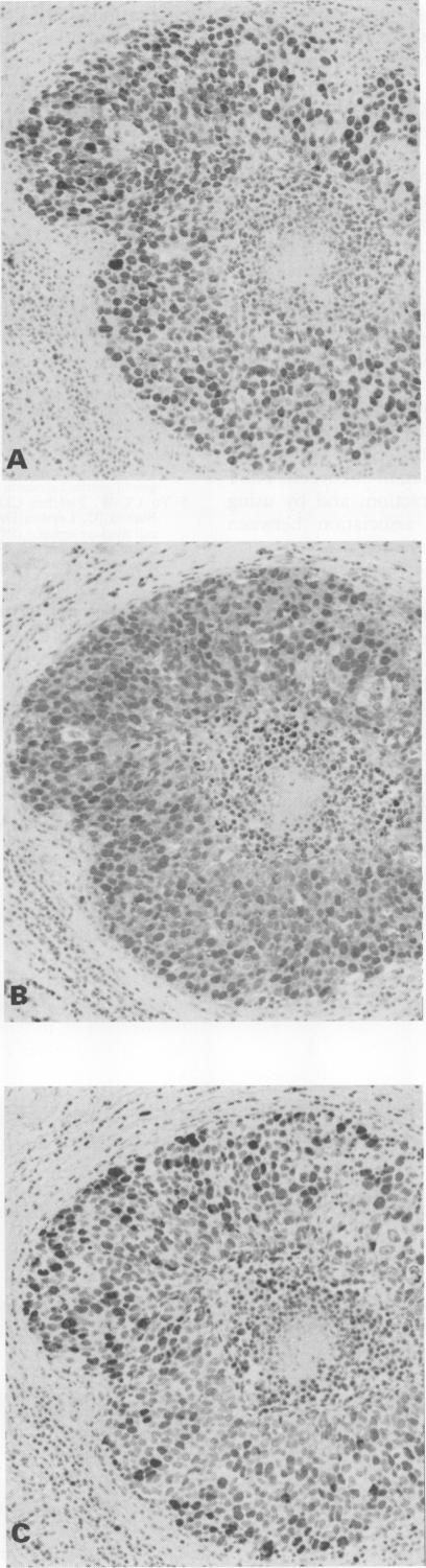
The staining patterns obtained with two antibodies against proliferating cell nuclear antigen (PC10 and 19A2) and another cell cycle associated antibody (KiS1) were compared with each other and with a number of established prognostic markers of breast carcinoma. Although PC10 and 19A2 staining patterns were similar, only the latter was significantly associated with KiS1 antibody staining. These findings suggest that the two PCNA antibodies detect different epitopes. KiS1 was the only antibody to show an association with S phase fraction measured by flow cytometry (p < 0.001). It was also associated with histological grade (p = 0.003), oestrogen receptors (p = 0.045), and DNA index (p = 0.007). PC10 showed no association with any of the markers of prognosis, while 19A2 was associated with histological grade (p = 0.017) and oestrogen receptors (p = 0.043). The two PCNA antibodies do not seem to be of value in measuring proliferative activity nor do they seem to be associated with established markers of prognosis in breast cancer.

摘要

将两种抗增殖细胞核抗原的抗体(PC10和19A2)以及另一种细胞周期相关抗体(KiS1)的染色模式相互进行比较,并与一些已确立的乳腺癌预后标志物进行比较。尽管PC10和19A2的染色模式相似,但只有后者与KiS1抗体染色显著相关。这些发现表明,两种PCNA抗体检测的是不同的表位。KiS1是唯一一种与通过流式细胞术测量的S期分数相关的抗体(p < 0.001)。它还与组织学分级(p = 0.003)、雌激素受体(p = 0.045)和DNA指数(p = 0.007)相关。PC10与任何预后标志物均无关联,而19A2与组织学分级(p = 0.017)和雌激素受体(p = 0.043)相关。这两种PCNA抗体在测量增殖活性方面似乎没有价值,在乳腺癌中似乎也与已确立的预后标志物无关。

https://cdn.ncbi.nlm.nih.gov/pmc/blobs/ed34/501726/4f7060f9130b/jclinpath00213-0059-a.jpg

相似文献

1
Comparison of three cell cycle associated antigens as markers of proliferative activity and prognosis in breast carcinoma.三种细胞周期相关抗原作为乳腺癌增殖活性和预后标志物的比较
J Clin Pathol. 1993 Dec;46(12):1126-8. doi: 10.1136/jcp.46.12.1126.
2
Cell-cycle-related staining patterns of anti-proliferating cell nuclear antigen monoclonal antibodies. Comparison with BrdUrd labeling and Ki-67 staining.抗增殖细胞核抗原单克隆抗体的细胞周期相关染色模式。与溴脱氧尿苷标记和Ki-67染色的比较。
Am J Pathol. 1991 May;138(5):1165-72.
3
Estimation of tumor growth fractions in archival formalin-fixed, paraffin-embedded tissues using two anti-PCNA/Cyclin monoclonal antibodies. Factors affecting reactivity.使用两种抗增殖细胞核抗原/细胞周期蛋白单克隆抗体对存档福尔马林固定石蜡包埋组织中的肿瘤生长分数进行评估。影响反应性的因素。
Am J Pathol. 1992 Dec;141(6):1453-8.
4
Proliferating cell nuclear antigen immunohistochemistry using monoclonal antibody 19A2 and a new antigen retrieval technique has prognostic impact in archival paraffin-embedded node-negative breast cancer.使用单克隆抗体19A2的增殖细胞核抗原免疫组织化学和一种新的抗原修复技术对存档石蜡包埋的淋巴结阴性乳腺癌具有预后影响。
Am J Pathol. 1993 Apr;142(4):1081-9.
5
The assessment of proliferating cell nuclear antigen (PCNA) immunostaining in primary gastrointestinal lymphomas and its relationship to histological grade, S+G2+M phase fraction (flow cytometric analysis) and prognosis.原发性胃肠道淋巴瘤中增殖细胞核抗原(PCNA)免疫染色的评估及其与组织学分级、S+G2+M期细胞比例(流式细胞术分析)和预后的关系。
Histopathology. 1991 Jul;19(1):21-7. doi: 10.1111/j.1365-2559.1991.tb00890.x.
6
Proliferating-cell nuclear antigen (PC10) immunolabelling and other proliferation indices as prognostic factors in breast cancer.增殖细胞核抗原(PC10)免疫标记及其他增殖指标作为乳腺癌的预后因素
J Cancer Res Clin Oncol. 1993;119(5):288-94. doi: 10.1007/BF01212727.
7
Assessment of proliferating cell nuclear antigen (PCNA) in breast cancer using anti-PCNA PC10 and 19A2: correlation with 5-bromo-2'-deoxyuridine or tritiated thymidine labeling and flow cytometric analysis.使用抗增殖细胞核抗原(PCNA)的PC10和19A2抗体评估乳腺癌中的增殖细胞核抗原:与5-溴-2'-脱氧尿苷或氚标记胸腺嘧啶核苷标记及流式细胞术分析的相关性
Biotech Histochem. 1994 Jul;69(4):203-12. doi: 10.3109/10520299409106288.
8
Minimal breast cancer: analysis of proliferative activity using a monoclonal antibody to proliferating cell nuclear antigen (PCNA) and its relationship to histological grade and prognosis.微小乳腺癌:应用抗增殖细胞核抗原(PCNA)单克隆抗体分析增殖活性及其与组织学分级和预后的关系。
Pathologica. 1994 Apr;86(2):150-6.
9
Proliferative activity determined by DNA flow cytometry and proliferating cell nuclear antigen (PCNA) immunohistochemistry as a prognostic factor in prostatic carcinoma.通过DNA流式细胞术和增殖细胞核抗原(PCNA)免疫组织化学测定的增殖活性作为前列腺癌的预后因素。
J Pathol. 1992 Sep;168(1):7-13. doi: 10.1002/path.1711680103.
10
Cell proliferation in breast carcinoma assessed by a PCNA grading system and its relation to other prognostic variables.
Surg Oncol. 1993;2(1):59-63. doi: 10.1016/0960-7404(93)90045-z.

引用本文的文献

1
Evaluation of ER-α, ER-Β1 and ER-Β2 expression and correlation with clinicopathologic factors in invasive luminal subtype breast cancers.浸润性腔面型乳腺癌中 ER-α、ER-Β1 和 ER-Β2 的表达及其与临床病理因素的相关性评价。
Clin Transl Oncol. 2012 Mar;14(3):225-31. doi: 10.1007/s12094-012-0788-0.
2
Determination of cell proliferation.细胞增殖的测定
Clin Mol Pathol. 1995 Feb;48(1):M2-5. doi: 10.1136/mp.48.1.m2.
3
Prognostic significance of the proliferative activity in neuroblastoma.神经母细胞瘤增殖活性的预后意义

本文引用的文献

1
Ki-S1, a novel proliferative marker: flow cytometric assessment of staining in human breast carcinoma cells.Ki-S1,一种新型增殖标志物:人乳腺癌细胞染色的流式细胞术评估
Br J Cancer. 1993 Apr;67(4):657-62. doi: 10.1038/bjc.1993.122.
2
Correlation of proliferative activity in breast tissue using PCNA/cyclin.使用增殖细胞核抗原/细胞周期蛋白对乳腺组织中的增殖活性进行相关性分析。
Hum Pathol. 1990 Jul;21(7):781. doi: 10.1016/0046-8177(90)90039-8.
3
The assessment of proliferating cell nuclear antigen (PCNA) immunostaining in primary gastrointestinal lymphomas and its relationship to histological grade, S+G2+M phase fraction (flow cytometric analysis) and prognosis.
Am J Pathol. 1997 Jan;150(1):133-45.
4
Lack of prognostic significance of the monoclonal antibody Ki-S1, a novel marker of proliferative activity, in node-negative breast carcinoma.单克隆抗体Ki-S1作为增殖活性的一种新标志物,在淋巴结阴性乳腺癌中缺乏预后意义。
Breast Cancer Res Treat. 1996;37(2):123-33. doi: 10.1007/BF01806494.
5
Cell proliferation in prostatic carcinoma: comparative analysis of Ki-67, MIB-1 and PCNA.前列腺癌中的细胞增殖:Ki-67、MIB-1和增殖细胞核抗原的比较分析
Histochem J. 1995 Mar;27(3):196-203.
6
Differentiation of short-wavelength-sensitive cones by NADPH diaphorase histochemistry.通过NADPH黄递酶组织化学对短波敏感视锥细胞进行分化。
Proc Natl Acad Sci U S A. 1995 May 23;92(11):5121-3. doi: 10.1073/pnas.92.11.5121.
7
Proliferation-associated nuclear antigen Ki-S1 is identical with topoisomerase II alpha. Delineation of a carboxy-terminal epitope with peptide antibodies.增殖相关核抗原Ki-S1与拓扑异构酶IIα相同。用肽抗体描绘羧基末端表位。
Am J Pathol. 1995 Jun;146(6):1302-8.
原发性胃肠道淋巴瘤中增殖细胞核抗原(PCNA)免疫染色的评估及其与组织学分级、S+G2+M期细胞比例(流式细胞术分析)和预后的关系。
Histopathology. 1991 Jul;19(1):21-7. doi: 10.1111/j.1365-2559.1991.tb00890.x.
4
Prognostic value of proliferating cell nuclear antigen in gastric carcinoma.增殖细胞核抗原在胃癌中的预后价值
J Clin Pathol. 1991 Aug;44(8):655-9. doi: 10.1136/jcp.44.8.655.
5
KiS1--a novel monoclonal antibody which recognizes proliferating cells: evaluation of its relationship to prognosis in mammary carcinoma.
J Pathol. 1992 Oct;168(2):179-85. doi: 10.1002/path.1711680205.
6
PC-10 antibody to proliferating cell nuclear antigen (PCNA) is not related to prognosis in human breast carcinoma.针对增殖细胞核抗原(PCNA)的PC-10抗体与人类乳腺癌的预后无关。
Growth Regul. 1992 Dec;2(4):145-50.
7
Flow cytometric characterisation of proliferating cell nuclear antigen using the monoclonal antibody PC10.使用单克隆抗体PC10对增殖细胞核抗原进行流式细胞术表征。
Eur J Cancer. 1992;28A(12):2010-7. doi: 10.1016/0959-8049(92)90250-6.
8
PCNA and Ki67 expression in breast carcinoma: correlations with clinical and biological variables.
J Clin Pathol. 1992 May;45(5):416-9. doi: 10.1136/jcp.45.5.416.
9
A comparison of proliferating cell nuclear antigen (PCNA) immunostaining, nucleolar organizer region (AgNOR) staining, and histological grading in gastrointestinal stromal tumours.胃肠道间质瘤中增殖细胞核抗原(PCNA)免疫染色、核仁组成区(AgNOR)染色及组织学分级的比较
J Pathol. 1992 Feb;166(2):147-52. doi: 10.1002/path.1711660210.