Veale D J, Barnes L, Rogers S, FitzGerald O
St Vincent's Hospital, Department of Rheumatology, Dublin, Ireland.
Ann Rheum Dis. 1994 Jul;53(7):450-4. doi: 10.1136/ard.53.7.450.
To examine the immunohistological features in the involved skin of patients with psoriatic arthritis (PA) (n = 15), compared with those in involved skin from patients with psoriasis but no arthritis (n = 5), and with a group with normal skin (n = 4), to identify markers for arthritis in psoriasis.
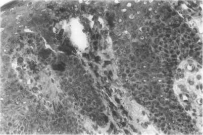
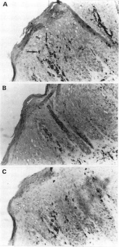
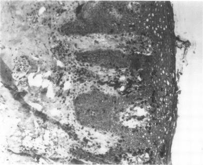
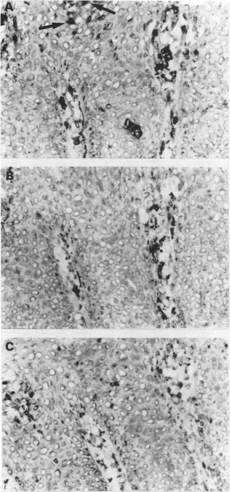
Skin was obtained from patients by 6 mm punch biopsy and normal skin was provided by the department of plastic surgery. Samples were stained with monoclonal antibodies against T cells (CD3, CD8, CD4, CD45Ro), B cells (CD20), macrophages (mac387), vascular endothelium (FVIII-related antigen) and a Langerhan's cell marker (p155). The number of cells/vessels staining with each monoclonal antibody was calculated and serial sections of skin were examined to estimate the presence of DR+ keratinocytes.
There were significantly more CD45Ro T-cells and blood vessels in patients with psoriatic arthritis compared with both psoriasis alone, and with normal controls (p < 0.02). While B-cells were not seen in psoriasis without arthritis or in normal skin, a small but significant number were observed in PA (p < 0.02). Furthermore, while DR+ keratinocytes were present in both psoriatic arthritis and psoriasis skin, there were significantly more DR+ cells in the psoriatic arthritis epidermis compared with psoriasis alone (p < 0.02).
This study suggests that increased numbers of CD45Ro T-cells, greater vascularity, the presence of B-cells, and increased numbers of DR+ epidermal cells are markers for arthritis in patients with psoriasis.
与无关节炎的银屑病患者(n = 5)的受累皮肤以及正常皮肤组(n = 4)相比,研究银屑病关节炎(PA)患者(n = 15)受累皮肤的免疫组织学特征,以确定银屑病中关节炎的标志物。
通过6毫米打孔活检从患者获取皮肤,正常皮肤由整形外科提供。样本用抗T细胞(CD3、CD8、CD4、CD45Ro)、B细胞(CD20)、巨噬细胞(mac387)、血管内皮(FVIII相关抗原)和朗格汉斯细胞标志物(p155)的单克隆抗体染色。计算每种单克隆抗体染色的细胞/血管数量,并检查皮肤连续切片以评估DR +角质形成细胞的存在情况。
与单独的银屑病患者和正常对照组相比,银屑病关节炎患者的CD45Ro T细胞和血管明显更多(p < 0.02)。在无关节炎的银屑病患者或正常皮肤中未见到B细胞,但在银屑病关节炎患者中观察到少量但显著数量的B细胞(p < 0.02)。此外,虽然DR +角质形成细胞存在于银屑病关节炎和银屑病皮肤中,但与单独的银屑病相比,银屑病关节炎表皮中的DR +细胞明显更多(p < 0.02)。
本研究表明,CD45Ro T细胞数量增加、血管增多、B细胞的存在以及DR +表皮细胞数量增加是银屑病患者关节炎的标志物。