• 文献检索
  • 文档翻译
  • 深度研究
  • 学术资讯
  • Suppr Zotero 插件Zotero 插件
  • 邀请有礼
  • 套餐&价格
  • 历史记录
应用&插件
Suppr Zotero 插件Zotero 插件浏览器插件Mac 客户端Windows 客户端微信小程序
定价
高级版会员购买积分包购买API积分包
服务
文献检索文档翻译深度研究API 文档MCP 服务
关于我们
关于 Suppr公司介绍联系我们用户协议隐私条款
关注我们

Suppr 超能文献

核心技术专利:CN118964589B侵权必究
粤ICP备2023148730 号-1Suppr @ 2026

文献检索

告别复杂PubMed语法,用中文像聊天一样搜索,搜遍4000万医学文献。AI智能推荐,让科研检索更轻松。

立即免费搜索

文件翻译

保留排版,准确专业,支持PDF/Word/PPT等文件格式,支持 12+语言互译。

免费翻译文档

深度研究

AI帮你快速写综述,25分钟生成高质量综述,智能提取关键信息,辅助科研写作。

立即免费体验

Mesangial cell apoptosis: the major mechanism for resolution of glomerular hypercellularity in experimental mesangial proliferative nephritis.

作者信息

Baker A J, Mooney A, Hughes J, Lombardi D, Johnson R J, Savill J

机构信息

Department of Medicine, University Hospital, Nottingham, United Kingdom.

出版信息

J Clin Invest. 1994 Nov;94(5):2105-16. doi: 10.1172/JCI117565.

DOI:10.1172/JCI117565
PMID:7962557
原文链接:https://pmc.ncbi.nlm.nih.gov/articles/PMC294654/
Abstract

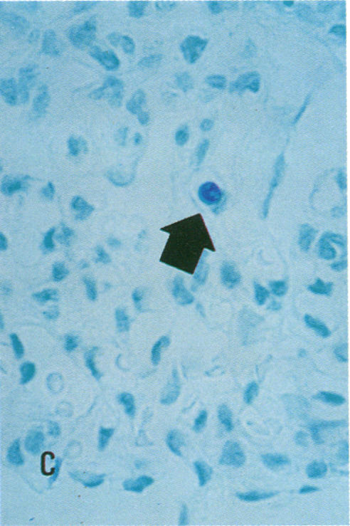
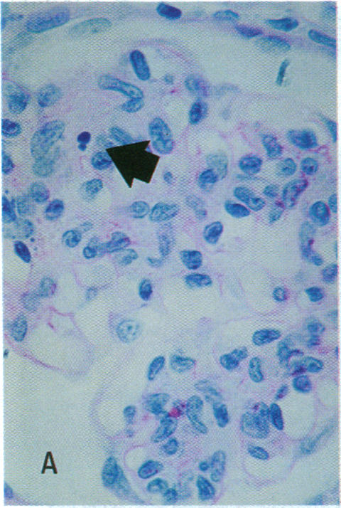
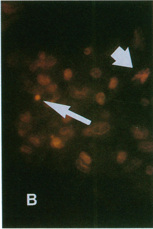
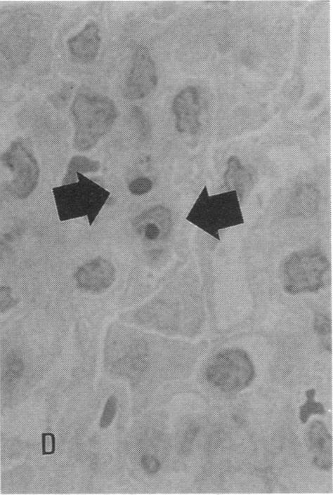
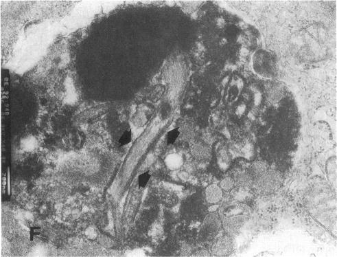
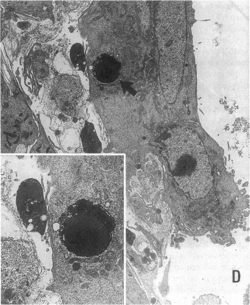
Increases in mesangial cell number may herald glomerular scarring, but they are not irreversible. This study sought mechanisms by which surplus glomerular mesangial cells can be cleared. A small proportion of cultured mesangial cells exhibited typical morphological features of apoptosis (programmed cell death), which was increased by growth factor deprivation or exposure to cycloheximide, stimuli known to increase apoptosis in other cell types. Apoptosis was confirmed by typical internucleosomal chromatin cleavage. In vivo, clear morphological evidence of mesangial apoptosis leading to phagocytosis by neighboring mesangial cells was obtained in self-limited mesangial proliferation induced in rats by Thy1.1 antibody, apoptosis occurring approximately 10-fold more frequently than in the healthy rat glomerulus. Indeed, changes in glomerular cell number in Thy1.1 nephritis strongly suggested that apoptosis is the major cell clearance mechanism counterbalancing cell division, thereby mediating resolution of glomerular hypercellularity in experimental mesangial proliferation.

摘要
https://cdn.ncbi.nlm.nih.gov/pmc/blobs/dd0d/294654/0658585b69cb/jcinvest00036-0409-a.jpg
https://cdn.ncbi.nlm.nih.gov/pmc/blobs/dd0d/294654/a25521eab52c/jcinvest00036-0405-a.jpg
https://cdn.ncbi.nlm.nih.gov/pmc/blobs/dd0d/294654/6e59895376a2/jcinvest00036-0405-b.jpg
https://cdn.ncbi.nlm.nih.gov/pmc/blobs/dd0d/294654/b1e9f4d198a1/jcinvest00036-0405-c.jpg
https://cdn.ncbi.nlm.nih.gov/pmc/blobs/dd0d/294654/8fe27782e99c/jcinvest00036-0405-d.jpg
https://cdn.ncbi.nlm.nih.gov/pmc/blobs/dd0d/294654/20f6f6d78685/jcinvest00036-0405-e.jpg
https://cdn.ncbi.nlm.nih.gov/pmc/blobs/dd0d/294654/cc39a9bde870/jcinvest00036-0405-f.jpg
https://cdn.ncbi.nlm.nih.gov/pmc/blobs/dd0d/294654/31b201366aac/jcinvest00036-0405-g.jpg
https://cdn.ncbi.nlm.nih.gov/pmc/blobs/dd0d/294654/b7910085dff4/jcinvest00036-0406-a.jpg
https://cdn.ncbi.nlm.nih.gov/pmc/blobs/dd0d/294654/66b639046250/jcinvest00036-0406-b.jpg
https://cdn.ncbi.nlm.nih.gov/pmc/blobs/dd0d/294654/1dcc24a0be81/jcinvest00036-0407-a.jpg
https://cdn.ncbi.nlm.nih.gov/pmc/blobs/dd0d/294654/7f3f4bbdc262/jcinvest00036-0408-a.jpg
https://cdn.ncbi.nlm.nih.gov/pmc/blobs/dd0d/294654/eb9fd8e9bc22/jcinvest00036-0408-b.jpg
https://cdn.ncbi.nlm.nih.gov/pmc/blobs/dd0d/294654/8ba225dacb87/jcinvest00036-0408-c.jpg
https://cdn.ncbi.nlm.nih.gov/pmc/blobs/dd0d/294654/3f63d2771a46/jcinvest00036-0408-d.jpg
https://cdn.ncbi.nlm.nih.gov/pmc/blobs/dd0d/294654/0658585b69cb/jcinvest00036-0409-a.jpg

相似文献

1
Mesangial cell apoptosis: the major mechanism for resolution of glomerular hypercellularity in experimental mesangial proliferative nephritis.
J Clin Invest. 1994 Nov;94(5):2105-16. doi: 10.1172/JCI117565.
2
P2 receptor antagonist PPADS inhibits mesangial cell proliferation in experimental mesangial proliferative glomerulonephritis.P2受体拮抗剂PPADS可抑制实验性系膜增生性肾小球肾炎中系膜细胞的增殖。
Kidney Int. 2002 Nov;62(5):1659-71. doi: 10.1046/j.1523-1755.2002.00621.x.
3
Effects of 1,25(OH)2D3 in experimental mesangial proliferative nephritis in rats.1,25-二羟维生素D3对大鼠实验性系膜增生性肾炎的影响
Kidney Int. 2001 Jul;60(1):87-95. doi: 10.1046/j.1523-1755.2001.00775.x.
4
SPARC is expressed by mesangial cells in experimental mesangial proliferative nephritis and inhibits platelet-derived-growth-factor-medicated mesangial cell proliferation in vitro.在实验性系膜增生性肾炎中,系膜细胞表达SPARC,并在体外抑制血小板衍生生长因子介导的系膜细胞增殖。
Am J Pathol. 1996 Apr;148(4):1153-67.
5
Methylprednisolone accelerates the resolution of glomerulonephritis by sensitizing mesangial cells to apoptosis.
Exp Nephrol. 2001;9(5):317-26. doi: 10.1159/000052627.
6
Pentoxifylline attenuates experimental mesangial proliferative glomerulonephritis.己酮可可碱可减轻实验性系膜增生性肾小球肾炎。
Kidney Int. 1999 Sep;56(3):932-43. doi: 10.1046/j.1523-1755.1999.00636.x.
7
Role of interleukin-1 in mesangial cell proliferation and matrix deposition in experimental mesangioproliferative nephritis.白细胞介素-1在实验性系膜增生性肾炎中系膜细胞增殖和基质沉积中的作用
Am J Pathol. 1997 Jul;151(1):141-50.
8
Apoptosis in progressive crescentic glomerulonephritis.进行性新月体性肾小球肾炎中的细胞凋亡
Lab Invest. 1996 May;74(5):941-51.
9
Complement-mediated killing of mesangial cells in experimental glomerulonephritis: cell death by a combination of apoptosis and necrosis.补体介导的实验性肾小球肾炎中系膜细胞杀伤作用:凋亡与坏死相结合导致的细胞死亡
Nephron. 2000 Oct;86(2):152-60. doi: 10.1159/000045734.
10
Endothelial progenitor cell-derived extracellular vesicles protect from complement-mediated mesangial injury in experimental anti-Thy1.1 glomerulonephritis.内皮祖细胞衍生的细胞外囊泡可保护实验性抗Thy1.1肾小球肾炎免受补体介导的系膜损伤。
Nephrol Dial Transplant. 2015 Mar;30(3):410-22. doi: 10.1093/ndt/gfu364. Epub 2014 Dec 8.

引用本文的文献

1
Integrin Alpha8 Beta1 (81): An In-Depth Review of an Overlooked RGD-Binding Receptor.整合素α8β1(81):对一个被忽视的RGD结合受体的深入综述
Biocell. 2025;49(5):789-811. doi: 10.32604/biocell.2025.062325. Epub 2025 May 27.
2
Monocytes/Macrophages Promote Regional Immune Injury in Mesangial Proliferative Glomerulonephritis through Crosstalk with Activated Mesangial Cells.单核细胞/巨噬细胞通过与活化的系膜细胞相互作用促进系膜增生性肾小球肾炎的局部免疫损伤。
Research (Wash D C). 2025 Jun 2;8:0716. doi: 10.34133/research.0716. eCollection 2025.
3
Porphyromonas gingivalis-derived lipopolysaccharide promotes mesangial cell fibrosis via transforming growth factor-beta1/Smad signaling pathway in high glucose.

本文引用的文献

1
Trafficking of inflammatory macrophages from the kidney to draining lymph nodes during experimental glomerulonephritis.实验性肾小球肾炎期间炎性巨噬细胞从肾脏向引流淋巴结的迁移
Clin Exp Immunol. 1993 May;92(2):336-41. doi: 10.1111/j.1365-2249.1993.tb03401.x.
2
Heparin suppresses mesangial cell proliferation and matrix expansion in experimental mesangioproliferative glomerulonephritis.肝素可抑制实验性系膜增生性肾小球肾炎中系膜细胞的增殖和基质扩张。
Kidney Int. 1993 Feb;43(2):369-80. doi: 10.1038/ki.1993.55.
3
Growth factors in glomerulonephritis.
牙龈卟啉单胞菌衍生的脂多糖通过高糖环境下的转化生长因子-β1/Smad信号通路促进系膜细胞纤维化。
J Dent Sci. 2025 Apr;20(2):989-994. doi: 10.1016/j.jds.2024.10.029. Epub 2024 Nov 21.
4
Mesangial cell-derived CircRNAs in chronic glomerulonephritis: RNA sequencing and bioinformatics analysis.肾小球肾炎中系膜细胞衍生的 CircRNAs:RNA 测序和生物信息学分析。
Ren Fail. 2024 Dec;46(2):2371059. doi: 10.1080/0886022X.2024.2371059. Epub 2024 Jul 1.
5
Macrophages communicate with mesangial cells through the CXCL12/DPP4 axis in lupus nephritis pathogenesis.巨噬细胞通过 CXCL12/DPP4 轴在狼疮性肾炎发病机制中与系膜细胞通讯。
Cell Death Dis. 2024 May 18;15(5):344. doi: 10.1038/s41419-024-06708-4.
6
Protective Effects of Rifampicin and Its Analog Rifampicin Quinone in a Mouse Model of Obesity-Induced Type 2 Diabetes.利福平及其类似物醌式利福平在肥胖诱导的2型糖尿病小鼠模型中的保护作用
ACS Pharmacol Transl Sci. 2023 Jan 12;6(2):253-269. doi: 10.1021/acsptsci.2c00082. eCollection 2023 Feb 10.
7
Mesangial cell: A hub in lupus nephritis.系膜细胞:狼疮肾炎的中心环节。
Front Immunol. 2022 Dec 14;13:1063497. doi: 10.3389/fimmu.2022.1063497. eCollection 2022.
8
Protective Effects of Naringenin from (var. Valencia) Peels against CCl-Induced Hepatic and Renal Injuries in Rats Assessed by Metabolomics, Histological and Biochemical Analyses.柚皮苷(橘红)对 CCl4 诱导的大鼠肝肾功能损伤的保护作用:基于代谢组学、组织学和生化分析的研究。
Nutrients. 2022 Feb 17;14(4):841. doi: 10.3390/nu14040841.
9
The Glomerulus According to the Mesangium.根据系膜的肾小球。
Front Med (Lausanne). 2022 Jan 26;8:740527. doi: 10.3389/fmed.2021.740527. eCollection 2021.
10
Doxorubicin-Induced Fetal Mesangial Cell Death Occurs Independently of TRPC6 Channel Upregulation but Involves Mitochondrial Generation of Reactive Oxygen Species.多柔比星诱导的胎儿肾小球系膜细胞死亡不依赖于 TRPC6 通道上调,但涉及线粒体产生活性氧。
Int J Mol Sci. 2021 Jul 15;22(14):7589. doi: 10.3390/ijms22147589.
肾小球肾炎中的生长因子
Kidney Int. 1993 Jan;43(1):252-67. doi: 10.1038/ki.1993.39.
4
Transient elevations of cytosolic free calcium retard subsequent apoptosis in neutrophils in vitro.
J Clin Invest. 1993 Jul;92(1):446-55. doi: 10.1172/JCI116587.
5
Role of mesenchymal cell death in lung remodeling after injury.间充质细胞死亡在损伤后肺重塑中的作用。
J Clin Invest. 1993 Jul;92(1):388-97. doi: 10.1172/JCI116578.
6
Glomerulosclerosis induced by in vivo transfection of transforming growth factor-beta or platelet-derived growth factor gene into the rat kidney.通过将转化生长因子-β或血小板衍生生长因子基因体内转染至大鼠肾脏所诱导的肾小球硬化。
J Clin Invest. 1993 Dec;92(6):2597-601. doi: 10.1172/JCI116874.
7
Molecular regulation of apoptosis: genetic controls on cell death.细胞凋亡的分子调控:细胞死亡的基因控制
Cell. 1993 Sep 10;74(5):777-9. doi: 10.1016/0092-8674(93)90457-2.
8
Large-scale normal cell death in the developing rat kidney and its reduction by epidermal growth factor.发育中大鼠肾脏的大规模正常细胞死亡及其被表皮生长因子的减少。
Development. 1993 Jul;118(3):777-84. doi: 10.1242/dev.118.3.777.
9
Cell death: the significance of apoptosis.细胞死亡:细胞凋亡的意义
Int Rev Cytol. 1980;68:251-306. doi: 10.1016/s0074-7696(08)62312-8.
10
Cell renewal of glomerular cell types in normal rats. An autoradiographic analysis.正常大鼠肾小球细胞类型的细胞更新。放射自显影分析。
Kidney Int. 1983 Nov;24(5):626-31. doi: 10.1038/ki.1983.203.