Nyberg S L, Remmel R P, Mann H J, Peshwa M V, Hu W S, Cerra F B
Department of Surgery, University of Minnesota, Minneapolis.
Ann Surg. 1994 Jul;220(1):59-67.
Metabolic activity of transformed human liver (Hep G2) cells and primary rat hepatocytes were compared during in vitro application of a gel entrapment bioartificial liver.
Clinical trials of bioartificial liver devices containing either transformed liver cells or primary hepatocytes have been initiated. A study comparing transformed liver cells and primary hepatocytes in a bioartificial liver under similar conditions has not been reported previously.
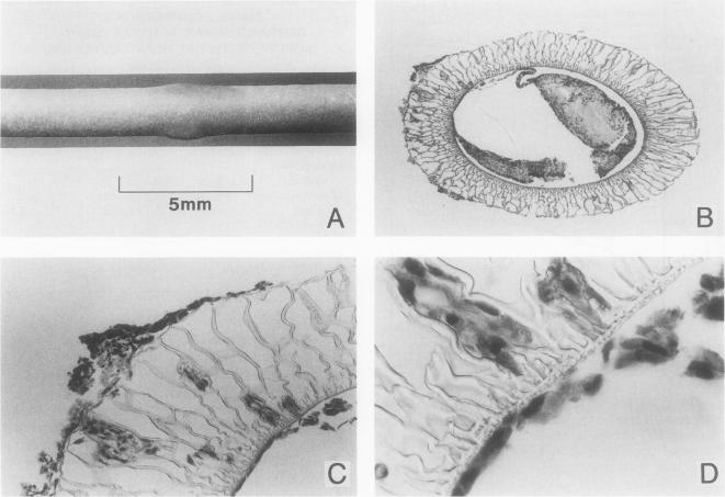
Gel entrapment bioartificial liver devices were inoculated with 100 million cells, Hep G2 cell line (n = 4), or rat hepatocytes (n = 16), and studied for up to 60 days of in vitro cultivation.
Hep G2 cells grew to confluence within the gel entrapment configuration with a doubling time of 20 +/- 3 hours. Rat hepatocytes significantly outperformed Hep G2 cells at confluence in all categories of biotransformation, including ureagenesis (3.5 +/- 0.7 vs. 0.3 +/- 0.1 mumol/hr, p < 0.05), glucuronidation (630 +/- 75 vs. 21 +/- 2 nmol/hr, p < 0.005), sulfation (59 +/- 13 vs. 5 +/- 2 nmol/hr, p < 0.05), and oxidation (233 +/- 38 vs. < 1 nmol/hr, p < 0.005). At the conclusion of one experiment, Hep G2 cells were found in the extracapillary compartment of the bioartificial liver, analogous to the patient's compartment during clinical application.
Primary rat hepatocytes were superior to the Hep G2 cell line as the source of hepatic function in a bioartificial liver and avoided the potential risk of tumor transmigration from the bioartificial liver into the patient's circulation.
在体外应用凝胶包封型生物人工肝期间,比较转化的人肝细胞(Hep G2)和原代大鼠肝细胞的代谢活性。
包含转化肝细胞或原代肝细胞的生物人工肝装置的临床试验已经启动。此前尚未有关于在相似条件下比较生物人工肝中转化肝细胞和原代肝细胞的研究报道。
将凝胶包封型生物人工肝装置接种1亿个细胞,Hep G2细胞系(n = 4)或大鼠肝细胞(n = 16),并进行长达60天的体外培养研究。
Hep G2细胞在凝胶包封结构内生长至汇合状态,倍增时间为20±3小时。在所有生物转化类别中,汇合时大鼠肝细胞的表现均显著优于Hep G2细胞,包括尿素生成(3.5±0.7对0.3±0.1 μmol/小时,p < 0.05)、葡萄糖醛酸化(630±75对21±2 nmol/小时,p < 0.005)、硫酸化(