Bernauer W, Watson P G, Daicker B, Lightman S
Moorfields Eye Hospital, London.
Br J Ophthalmol. 1994 May;78(5):381-5. doi: 10.1136/bjo.78.5.381.
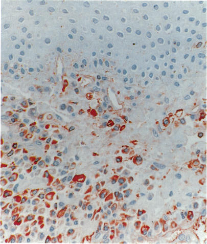
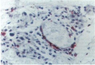
Scleritis can be a destructive disease frequently associated with autoimmune disorders. It is believed that primary vasculitis plays an important role in its pathogenesis, but little is known about the cellular effector mechanisms. The purpose of this study was to analyse the inflammatory cellular infiltrate in scleritis. Six episcleral biopsies and two enucleated eyes were studied. The episcleral biopsies were taken from patients with nodular scleritis. In one patient enucleation was done after perforation in anterior necrotising scleritis and, in the other after misdiagnosis of posterior scleritis as intraocular tumour. Morphological criteria and immunohistochemical methods were used to characterise the inflammatory cellular infiltrate. The inflammatory cells infiltrating the episcleral tissue were mainly T lymphocytes and macrophages. There was a predominance of CD4 positive cells, but only few lymphocytes were activated (expressed IL-2 receptor). The cells infiltrating the scleral fibres in the enucleated eyes consisted in both cases predominantly of T cells. Clusters of B cells were found in perivascular areas. In circumscribed areas neutrophils, macrophages, and plasma cells were part of the scleral infiltrate. Signs of a granulomatous process with activated macrophages (epithelioid and giant cells) were present in necrotising scleritis. Expression of major histocompatibility class II molecules (MHC II) was found on lymphocytes and rarely on macrophages. Signs of primary vasculitis were not found in any of the specimens. The cellular infiltrate in scleritis shows, at least at certain stages, features compatible with a T cell mediated (autoimmune) disorder, which may have major therapeutic implications.
巩膜炎是一种常与自身免疫性疾病相关的破坏性疾病。人们认为原发性血管炎在其发病机制中起重要作用,但对细胞效应机制了解甚少。本研究的目的是分析巩膜炎中的炎性细胞浸润情况。对6例巩膜活检组织和2只摘除眼球进行了研究。巩膜活检组织取自结节性巩膜炎患者。1例患者因前部坏死性巩膜炎穿孔后进行了眼球摘除,另1例因后巩膜炎被误诊为眼内肿瘤而进行了眼球摘除。采用形态学标准和免疫组化方法对炎性细胞浸润进行特征描述。浸润巩膜组织的炎性细胞主要是T淋巴细胞和巨噬细胞。CD4阳性细胞占优势,但只有少数淋巴细胞被激活(表达IL-2受体)。在两例摘除眼球中,浸润巩膜纤维的细胞主要由T细胞组成。在血管周围区域发现了B细胞簇。在局限性区域,中性粒细胞、巨噬细胞和浆细胞是巩膜浸润的一部分。坏死性巩膜炎中存在活化巨噬细胞(上皮样细胞和巨细胞)的肉芽肿形成过程的迹象。在淋巴细胞上发现了主要组织相容性复合体II类分子(MHC II)的表达,而在巨噬细胞上很少见。在任何标本中均未发现原发性血管炎的迹象。巩膜炎中的细胞浸润至少在某些阶段表现出与T细胞介导的(自身免疫性)疾病相符的特征,这可能具有重要的治疗意义。