• 文献检索
  • 文档翻译
  • 深度研究
  • 学术资讯
  • Suppr Zotero 插件Zotero 插件
  • 邀请有礼
  • 套餐&价格
  • 历史记录
应用&插件
Suppr Zotero 插件Zotero 插件浏览器插件Mac 客户端Windows 客户端微信小程序
定价
高级版会员购买积分包购买API积分包
服务
文献检索文档翻译深度研究API 文档MCP 服务
关于我们
关于 Suppr公司介绍联系我们用户协议隐私条款
关注我们

Suppr 超能文献

核心技术专利:CN118964589B侵权必究
粤ICP备2023148730 号-1Suppr @ 2026

文献检索

告别复杂PubMed语法,用中文像聊天一样搜索,搜遍4000万医学文献。AI智能推荐,让科研检索更轻松。

立即免费搜索

文件翻译

保留排版,准确专业,支持PDF/Word/PPT等文件格式,支持 12+语言互译。

免费翻译文档

深度研究

AI帮你快速写综述,25分钟生成高质量综述,智能提取关键信息,辅助科研写作。

立即免费体验

冠状病毒M蛋白在病毒粒子出芽位点之外的高尔基体复合物中积累。

Coronavirus M proteins accumulate in the Golgi complex beyond the site of virion budding.

作者信息

Klumperman J, Locker J K, Meijer A, Horzinek M C, Geuze H J, Rottier P J

机构信息

Department of Cell Biology, Utrecht University, The Netherlands.

出版信息

J Virol. 1994 Oct;68(10):6523-34. doi: 10.1128/JVI.68.10.6523-6534.1994.

DOI:10.1128/JVI.68.10.6523-6534.1994
PMID:8083990
原文链接:https://pmc.ncbi.nlm.nih.gov/articles/PMC237073/
Abstract

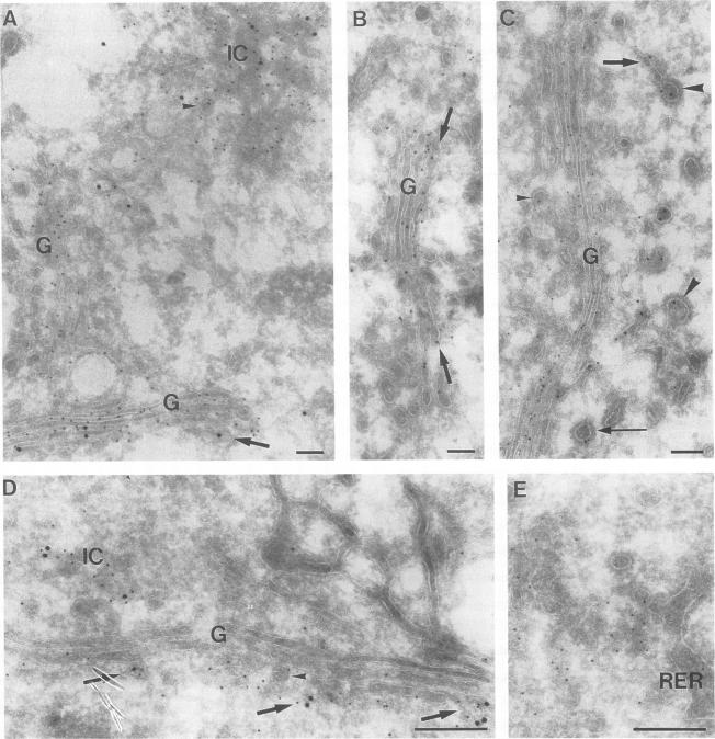
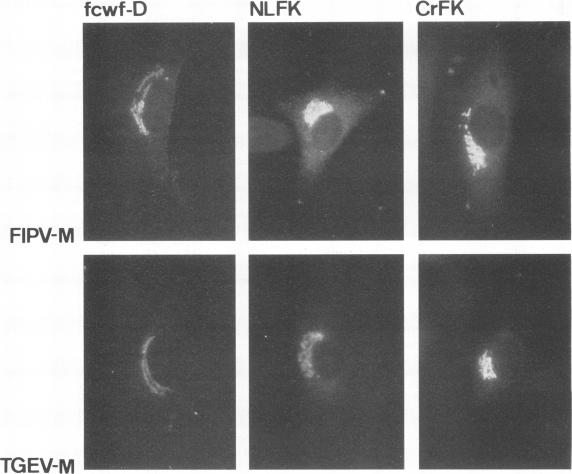
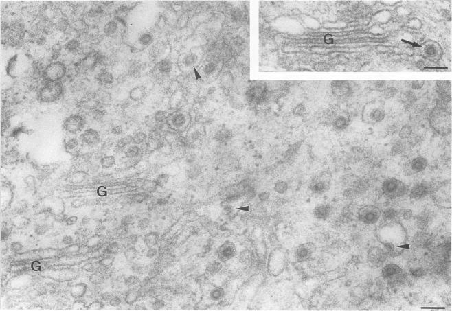
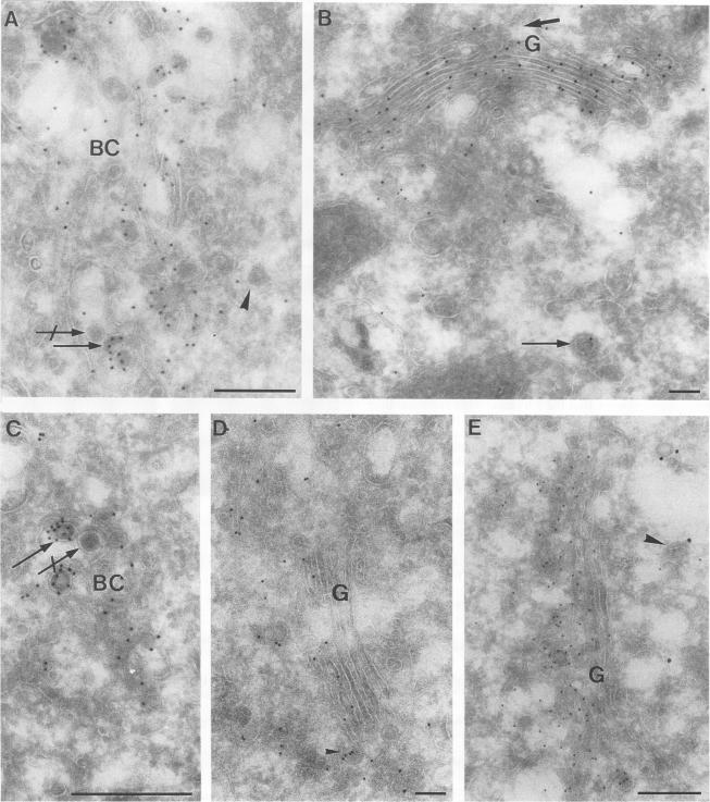
The prevailing hypothesis is that the intracellular site of budding of coronaviruses is determined by the localization of its membrane protein M (previously called E1). We tested this by analyzing the site of budding of four different coronaviruses in relation to the intracellular localization of their M proteins. Mouse hepatitis virus (MHV) and infectious bronchitis virus (IBV) grown in Sac(-) cells, and feline infectious peritonitis virus (FIPV) and transmissible gastroenteritis virus (TGEV) grown in CrFK cells, all budded exclusively into smooth-walled, tubulovesicular membranes located intermediately between the rough endoplasmic reticulum and Golgi complex, identical to the so-called budding compartment previously identified for MHV. Indirect immunofluorescence staining of the infected cells showed that all four M proteins accumulated in a perinuclear region. Immunogold microscopy localized MHV M and IBV M in the budding compartment; in addition, a dense labeling in the Golgi complex occurred, MHV M predominantly in trans-Golgi cisternae and trans-Golgi reticulum and IBV M mainly in the cis and medial Golgi cisternae. The corresponding M proteins of the four viruses, when independently expressed in a recombinant vaccinia virus system, also accumulated in the perinuclear area. Quantitative pulse-chase analysis of metabolically labeled cells showed that in each case the majority of the M glycoproteins carried oligosaccharide side chains with Golgi-specific modifications within 4 h after synthesis. Immunoelectron microscopy localized recombinant MHV M and IBV M to the same membranes as the respective proteins in coronavirus-infected cells, with the same cis-trans distribution over the Golgi complex. Our results demonstrate that some of the M proteins of the four viruses are transported beyond the budding compartment and are differentially retained by intrinsic retention signals; in addition to M, other viral and/or cellular factors are probably required to determine the site of budding.

摘要

目前流行的假说是冠状病毒出芽的细胞内位点由其膜蛋白M(以前称为E1)的定位决定。我们通过分析四种不同冠状病毒的出芽位点与其M蛋白的细胞内定位的关系来对此进行测试。在Sac(-)细胞中生长的小鼠肝炎病毒(MHV)和传染性支气管炎病毒(IBV),以及在CrFK细胞中生长的猫传染性腹膜炎病毒(FIPV)和猪传染性胃肠炎病毒(TGEV),均仅出芽到位于粗面内质网和高尔基体复合体之间的光滑壁、管状囊泡膜中,这与先前为MHV确定的所谓出芽区室相同。对感染细胞的间接免疫荧光染色显示,所有四种M蛋白都聚集在核周区域。免疫金显微镜观察将MHV M和IBV M定位在出芽区室中;此外,在高尔基体复合体中出现了密集标记,MHV M主要位于反式高尔基体潴泡和反式高尔基体网状结构中,而IBV M主要位于顺式和中间高尔基体潴泡中。这四种病毒的相应M蛋白在重组痘苗病毒系统中独立表达时,也聚集在核周区域。对代谢标记细胞的定量脉冲追踪分析表明,在每种情况下,大多数M糖蛋白在合成后4小时内携带具有高尔基体特异性修饰的寡糖侧链。免疫电子显微镜观察将重组MHV M和IBV M定位到与冠状病毒感染细胞中的相应蛋白相同的膜上,在高尔基体复合体上具有相同的顺反分布。我们的结果表明,这四种病毒的一些M蛋白被转运到出芽区室之外,并被内在的保留信号差异保留;除了M蛋白外,可能还需要其他病毒和/或细胞因子来决定出芽位点。

https://cdn.ncbi.nlm.nih.gov/pmc/blobs/cc8c/237073/f1a700a5ae4c/jvirol00019-0408-a.jpg
https://cdn.ncbi.nlm.nih.gov/pmc/blobs/cc8c/237073/54dd2fc3ed13/jvirol00019-0401-a.jpg
https://cdn.ncbi.nlm.nih.gov/pmc/blobs/cc8c/237073/11d7a44fcef8/jvirol00019-0402-a.jpg
https://cdn.ncbi.nlm.nih.gov/pmc/blobs/cc8c/237073/68cd25309749/jvirol00019-0403-a.jpg
https://cdn.ncbi.nlm.nih.gov/pmc/blobs/cc8c/237073/689edfabdbbd/jvirol00019-0404-a.jpg
https://cdn.ncbi.nlm.nih.gov/pmc/blobs/cc8c/237073/010e10020b03/jvirol00019-0404-b.jpg
https://cdn.ncbi.nlm.nih.gov/pmc/blobs/cc8c/237073/7c174d638161/jvirol00019-0405-a.jpg
https://cdn.ncbi.nlm.nih.gov/pmc/blobs/cc8c/237073/83832ef40cbf/jvirol00019-0406-a.jpg
https://cdn.ncbi.nlm.nih.gov/pmc/blobs/cc8c/237073/6413ce450841/jvirol00019-0407-a.jpg
https://cdn.ncbi.nlm.nih.gov/pmc/blobs/cc8c/237073/f1a700a5ae4c/jvirol00019-0408-a.jpg

相似文献

1
Coronavirus M proteins accumulate in the Golgi complex beyond the site of virion budding.冠状病毒M蛋白在病毒粒子出芽位点之外的高尔基体复合物中积累。
J Virol. 1994 Oct;68(10):6523-34. doi: 10.1128/JVI.68.10.6523-6534.1994.
2
Characterization of the budding compartment of mouse hepatitis virus: evidence that transport from the RER to the Golgi complex requires only one vesicular transport step.小鼠肝炎病毒出芽区室的特征:从内质网到高尔基体复合体的转运仅需一个囊泡转运步骤的证据。
J Cell Biol. 1994 Jan;124(1-2):55-70. doi: 10.1083/jcb.124.1.55.
3
Feline aminopeptidase N serves as a receptor for feline, canine, porcine, and human coronaviruses in serogroup I.猫氨基肽酶N作为血清I群中猫、犬、猪和人冠状病毒的受体。
J Virol. 1996 Dec;70(12):8669-74. doi: 10.1128/JVI.70.12.8669-8674.1996.
4
Replication of coronavirus MHV-A59 in sac- cells: determination of the first site of budding of progeny virions.冠状病毒MHV - A59在囊泡细胞中的复制:子代病毒粒子出芽的首个位点的确定。
Eur J Cell Biol. 1984 Mar;33(2):281-93.
5
Envelope glycoprotein interactions in coronavirus assembly.冠状病毒组装过程中的包膜糖蛋白相互作用。
J Cell Biol. 1995 Oct;131(2):339-49. doi: 10.1083/jcb.131.2.339.
6
Infection of AtT20 murine pituitary tumour cells by mouse hepatitis virus strain A59: virus budding is restricted to the Golgi region.A59株小鼠肝炎病毒对AtT20小鼠垂体瘤细胞的感染:病毒出芽仅限于高尔基体区域。
Eur J Cell Biol. 1985 May;37:203-12.
7
The cytoplasmic tail of mouse hepatitis virus M protein is essential but not sufficient for its retention in the Golgi complex.小鼠肝炎病毒M蛋白的胞质尾对于其滞留于高尔基体复合体至关重要,但并不充分。
J Biol Chem. 1994 Nov 11;269(45):28263-9.
8
Site of addition of N-acetyl-galactosamine to the E1 glycoprotein of mouse hepatitis virus-A59.N-乙酰半乳糖胺添加到小鼠肝炎病毒A59 E1糖蛋白的位点。
J Cell Biol. 1988 May;106(5):1475-87. doi: 10.1083/jcb.106.5.1475.
9
Structural maturation of the transmissible gastroenteritis coronavirus.传染性胃肠炎冠状病毒的结构成熟
J Virol. 1999 Oct;73(10):7952-64. doi: 10.1128/JVI.73.10.7952-7964.1999.
10
The ultrastructure of feline infectious peritonitis virus in feline embryonic lung cells.猫传染性腹膜炎病毒在猫胚胎肺细胞中的超微结构
J Gen Virol. 1982 Mar;59(Pt 1):23-8. doi: 10.1099/0022-1317-59-1-23.

引用本文的文献

1
Host protein ARF1 is a proviral factor for SARS-CoV-2 and a candidate broad-spectrum therapeutic target.宿主蛋白ARF1是SARS-CoV-2的一个前病毒因子和一个广谱治疗靶点候选物。
Nat Commun. 2025 Jul 9;16(1):6326. doi: 10.1038/s41467-025-61431-8.
2
Sequences in the Cytoplasmic Tail Contribute to the Intracellular Trafficking and the Cell Surface Localization of SARS-CoV-2 Spike Protein.细胞质尾段的序列有助于严重急性呼吸综合征冠状病毒2刺突蛋白的细胞内运输和细胞表面定位。
Biomolecules. 2025 Feb 14;15(2):280. doi: 10.3390/biom15020280.
3
Perturbation of lipogenesis hinders MERS-CoV assembly and release, but not the biogenesis of viral replication organelles.

本文引用的文献

1
Bidirectional membrane traffic between the endoplasmic reticulum and Golgi apparatus.内质网与高尔基体之间的双向膜泡运输
Trends Cell Biol. 1993 Mar;3(3):81-8. doi: 10.1016/0962-8924(93)90078-f.
2
AN ELECTRON MICROSCOPE STUDY OF THE DEVELOPMENT OF A MOUSE HEPATITIS VIRUS IN TISSUE CULTURE CELLS.小鼠肝炎病毒在组织培养细胞中发育的电子显微镜研究
J Cell Biol. 1965 Jan;24(1):57-78. doi: 10.1083/jcb.24.1.57.
3
Beta-COP localizes mainly to the cis-Golgi side in exocrine pancreas.β-COP主要定位于外分泌胰腺的顺式高尔基体一侧。
脂肪生成的扰动会阻碍中东呼吸综合征冠状病毒(MERS-CoV)的组装和释放,但不会影响病毒复制细胞器的生物发生。
J Virol. 2025 Mar 18;99(3):e0228224. doi: 10.1128/jvi.02282-24. Epub 2025 Feb 20.
4
The cytoplasmic tail of IBV spike mediates intracellular retention via interaction with COPI-coated vesicles in retrograde trafficking.传染性支气管炎病毒刺突蛋白的胞质尾通过与逆行转运中COPI包被囊泡相互作用介导细胞内滞留。
J Virol. 2025 Feb 25;99(2):e0216424. doi: 10.1128/jvi.02164-24. Epub 2025 Jan 22.
5
Coatomer complex I is required for the transport of SARS-CoV-2 progeny virions from the endoplasmic reticulum-Golgi intermediate compartment.COP-I衣被复合体对于严重急性呼吸综合征冠状病毒2(SARS-CoV-2)子代病毒粒子从内质网-高尔基体中间膜囊的转运是必需的。
mBio. 2025 Jan 8;16(1):e0333124. doi: 10.1128/mbio.03331-24. Epub 2024 Nov 29.
6
Mouse hepatitis virus JHMV I protein is required for maximal virulence.鼠肝炎病毒 JHMV I 蛋白是最大毒力所必需的。
J Virol. 2024 Sep 17;98(9):e0068024. doi: 10.1128/jvi.00680-24. Epub 2024 Aug 19.
7
Elucidating the behavior of the SARS-CoV-2 virus surface at vapor-liquid interfaces using molecular dynamics simulation.利用分子动力学模拟阐明 SARS-CoV-2 病毒表面在气液界面的行为。
Proc Natl Acad Sci U S A. 2024 Mar 26;121(13):e2317194121. doi: 10.1073/pnas.2317194121. Epub 2024 Mar 19.
8
Classification, replication, and transcription of ..的分类、复制和转录。 (你提供的原文不完整,请检查一下是否有遗漏内容)
Front Microbiol. 2024 Jan 24;14:1291761. doi: 10.3389/fmicb.2023.1291761. eCollection 2023.
9
Differential Ability of Spike Protein of SARS-CoV-2 Variants to Downregulate ACE2.SARS-CoV-2 变异株 Spike 蛋白下调 ACE2 的能力差异。
Int J Mol Sci. 2024 Jan 22;25(2):1353. doi: 10.3390/ijms25021353.
10
Membrane Condensation and Curvature Induced by SARS-CoV-2 Envelope Protein.由新冠病毒包膜蛋白诱导的膜凝聚和曲率
Langmuir. 2024 Feb 6;40(5):2646-2655. doi: 10.1021/acs.langmuir.3c03079. Epub 2024 Jan 22.
J Cell Biol. 1993 Apr;121(1):49-59. doi: 10.1083/jcb.121.1.49.
4
Retention of a cis Golgi protein requires polar residues on one face of a predicted alpha-helix in the transmembrane domain.顺式高尔基体蛋白的保留需要跨膜结构域中预测的α-螺旋一侧的极性残基。
Mol Biol Cell. 1993 Jul;4(7):695-704. doi: 10.1091/mbc.4.7.695.
5
Characterization of the budding compartment of mouse hepatitis virus: evidence that transport from the RER to the Golgi complex requires only one vesicular transport step.小鼠肝炎病毒出芽区室的特征:从内质网到高尔基体复合体的转运仅需一个囊泡转运步骤的证据。
J Cell Biol. 1994 Jan;124(1-2):55-70. doi: 10.1083/jcb.124.1.55.
6
Differences in the endosomal distributions of the two mannose 6-phosphate receptors.两种甘露糖6-磷酸受体在内体分布上的差异。
J Cell Biol. 1993 Jun;121(5):997-1010. doi: 10.1083/jcb.121.5.997.
7
Tunicamycin resistant glycosylation of coronavirus glycoprotein: demonstration of a novel type of viral glycoprotein.冠状病毒糖蛋白的衣霉素抗性糖基化:一种新型病毒糖蛋白的证明。
Virology. 1981 Dec;115(2):334-44. doi: 10.1016/0042-6822(81)90115-x.
8
Expression of feline infectious peritonitis coronavirus antigens on the surface of feline macrophage-like cells.
J Gen Virol. 1983 Sep;64 (Pt 9):1859-66. doi: 10.1099/0022-1317-64-9-1859.
9
Spectrophotometric quantitation of silver grains eluted from autoradiograms.从放射自显影片上洗脱的银颗粒的分光光度法定量
Anal Biochem. 1983 Sep;133(2):511-4. doi: 10.1016/0003-2697(83)90117-3.
10
Replication of coronavirus MHV-A59 in sac- cells: determination of the first site of budding of progeny virions.冠状病毒MHV - A59在囊泡细胞中的复制:子代病毒粒子出芽的首个位点的确定。
Eur J Cell Biol. 1984 Mar;33(2):281-93.