Wood A C, Waters C M, Garner A, Hickman J A
Cancer Research Campaign Molecular and Cellular Pharmacology Group, School of Biological Sciences, University of Manchester, UK.
Br J Cancer. 1994 Apr;69(4):663-9. doi: 10.1038/bjc.1994.128.
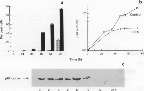
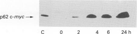
The kinetics of dexamethasone-induced death of CCRF CEM clone C7A human lymphoblastic leukaemia cells was determined with respect to changes in the expression of the c-myc protein. Cell death was characterised as being by apoptosis: cells with an intact plasma membrane had condensed chromatin and were characterised as having approximately 300 kbp fragments when DNA integrity was analysed by pulsed-field electrophoresis. Onset of apoptosis required a minimum of 36 h exposure to 5 microM dexamethasone; before this time no apoptotic cells were observed. This 36 h incubation period appeared to be necessary to prime the cells for subsequent death by apoptosis. In the continued presence of dexamethasone the percentage of apoptotic cells increased to 60% apoptotic cells by 54 h. Investigation of changes in c-myc protein showed that it was undetectable after 12 h of incubation with dexamethasone, although cells were not committed to die at this time. Cells were treated with dexamethasone for 54 h and for various pulsed periods with a non-toxic concentration of cycloheximide (200 nM). When cycloheximide was present during the first 36 h priming period of dexamethasone treatment, there was an immediate loss of c-myc protein and apoptosis at 54 h was completely inhibited. In contrast, there was no inhibition of apoptosis when dexamethasone-treated cells were incubated with an 18 h pulse of cycloheximide added after 36 h. Cells exposed to dexamethasone for 36 h ('primed') were given various periods of dexamethasone-free incubation before readdition of dexamethasone for a further 18 h. The longer the cells were free of drug after priming, the less susceptible they became to apoptosis, suggesting a slow decay of their 'memory' of the initial 36 h period of exposure. Cycloheximide inhibited the decay of this memory. Removal of dexamethasone after a 36 h exposure was characterised by a subsequent 24 h suppression of c-myc protein expression. Despite this, 90% of cells became refractory to apoptosis before the reappearance of c-myc protein. The evidence does not support the hypothesis that changes in c-myc expression are required for the engagement of apoptosis of CEM cells.
关于c-myc蛋白表达的变化,测定了地塞米松诱导CCRF CEM克隆C7A人淋巴细胞白血病细胞死亡的动力学。细胞死亡的特征为凋亡:当通过脉冲场电泳分析DNA完整性时,具有完整质膜的细胞染色质浓缩,其特征是具有约300 kbp的片段。凋亡的发生至少需要36小时暴露于5 microM地塞米松;在此之前未观察到凋亡细胞。这36小时的孵育期似乎是使细胞为随后的凋亡死亡做好准备所必需的。在地塞米松持续存在的情况下,到54小时时凋亡细胞的百分比增加到60%。对c-myc蛋白变化的研究表明,与地塞米松孵育12小时后检测不到该蛋白,尽管此时细胞尚未注定死亡。用54小时的地塞米松和无毒浓度的环己酰亚胺(200 nM)进行不同脉冲时间处理细胞。当地塞米松处理的最初36小时引发期存在环己酰亚胺时,c-myc蛋白立即丧失,54小时时凋亡被完全抑制。相反,当地塞米松处理的细胞在36小时后用18小时脉冲的环己酰亚胺孵育时,凋亡没有受到抑制。将暴露于地塞米松36小时(“引发”)的细胞在重新添加地塞米松再孵育18小时之前给予不同时间的无地塞米松孵育。引发后细胞无药物的时间越长,它们对凋亡的敏感性越低,这表明它们对最初36小时暴露的“记忆”在缓慢衰退。环己酰亚胺抑制了这种记忆的衰退。36小时暴露后去除地塞米松的特征是随后24小时c-myc蛋白表达受到抑制。尽管如此,90%的细胞在c-myc蛋白重新出现之前对凋亡变得不敏感。证据不支持c-myc表达变化是CEM细胞凋亡所必需的这一假设。