• 文献检索
  • 文档翻译
  • 深度研究
  • 学术资讯
  • Suppr Zotero 插件Zotero 插件
  • 邀请有礼
  • 套餐&价格
  • 历史记录
应用&插件
Suppr Zotero 插件Zotero 插件浏览器插件Mac 客户端Windows 客户端微信小程序
定价
高级版会员购买积分包购买API积分包
服务
文献检索文档翻译深度研究API 文档MCP 服务
关于我们
关于 Suppr公司介绍联系我们用户协议隐私条款
关注我们

Suppr 超能文献

核心技术专利:CN118964589B侵权必究
粤ICP备2023148730 号-1Suppr @ 2026

文献检索

告别复杂PubMed语法,用中文像聊天一样搜索,搜遍4000万医学文献。AI智能推荐,让科研检索更轻松。

立即免费搜索

文件翻译

保留排版,准确专业,支持PDF/Word/PPT等文件格式,支持 12+语言互译。

免费翻译文档

深度研究

AI帮你快速写综述,25分钟生成高质量综述,智能提取关键信息,辅助科研写作。

立即免费体验

上消化道神经内分泌肿瘤的定位

Localisation of neuroendocrine tumours of the upper gastrointestinal tract.

作者信息

Zimmer T, Ziegler K, Bäder M, Fett U, Hamm B, Riecken E O, Wiedenmann B

机构信息

Department of Internal Medicine, Steglitz Medical Centre, Free University of Berlin, Germany.

出版信息

Gut. 1994 Apr;35(4):471-5. doi: 10.1136/gut.35.4.471.

DOI:10.1136/gut.35.4.471
PMID:8174983
原文链接:https://pmc.ncbi.nlm.nih.gov/articles/PMC1374794/
Abstract

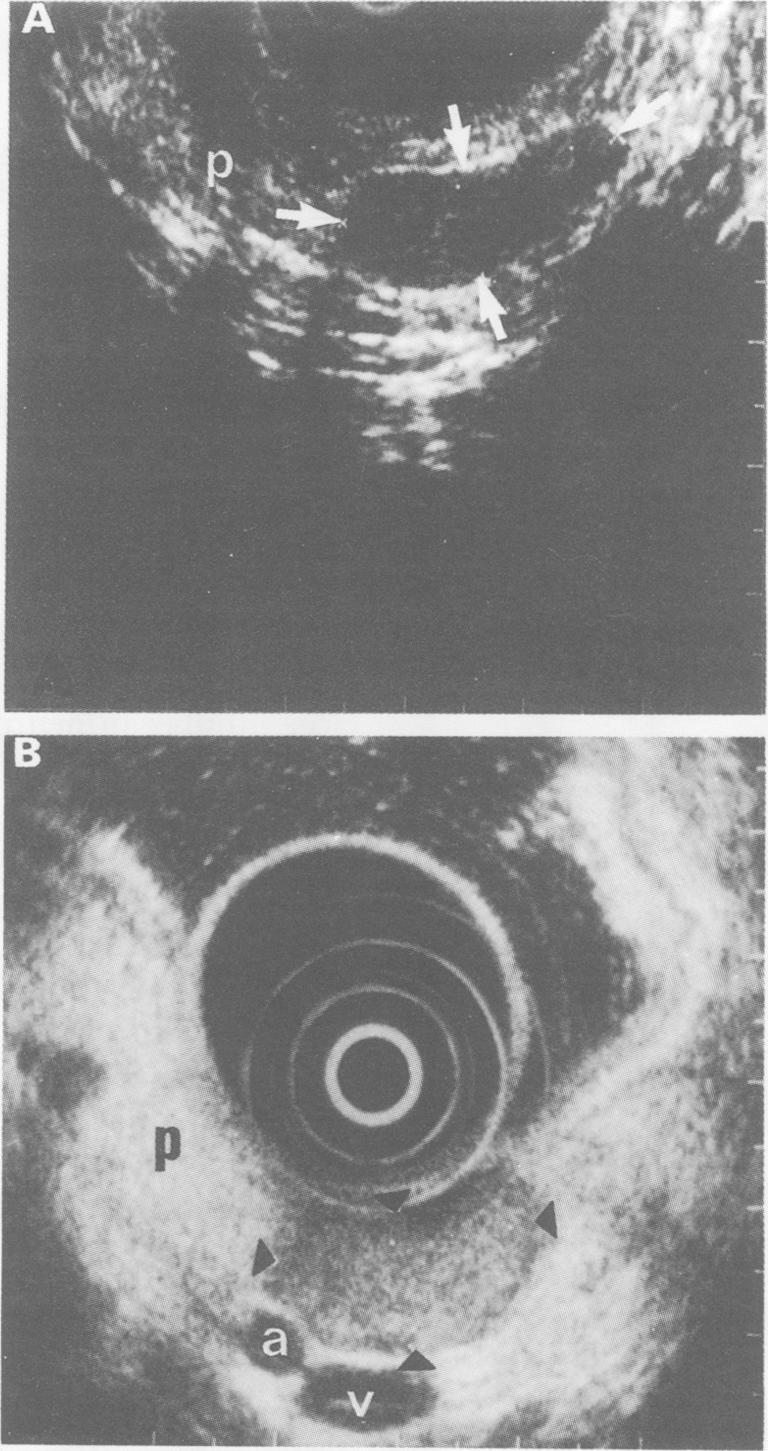
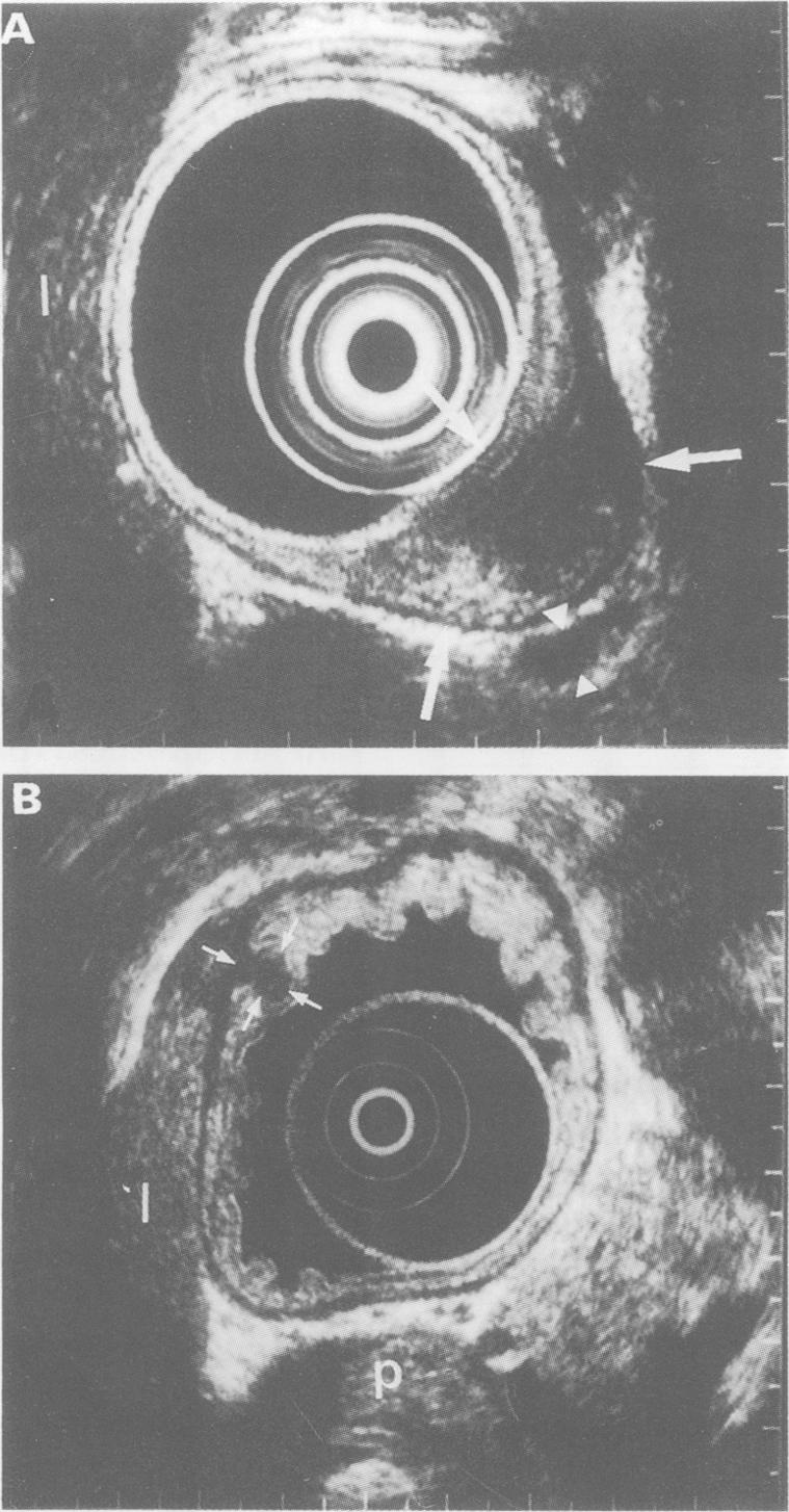
In order to localise neuroendocrine tumours of the foregut type (that is, of the stomach, duodenum, and pancreas), 18 patients were studied prospectively by endoscopic ultrasonography, computed tomography, transabdominal ultrasonography, magnetic resonance imaging, and somatostatin receptor scintigraphy. These 18 patients had a total of 25 primary tumour lesions which were verified histologically in tissue obtained by surgery or by ultrasound or endoscopy guided biopsy. Tumours were found in the stomach (n = 1), duodenum (n = 6), pancreas (n = 17), and liver (n = 1). Endoscopic ultrasonography had the highest sensitivity for tumour detection, followed by somatostatin receptor scintigraphy, computed tomography, transabdominal ultrasonography, and magnetic resonance imaging (88%, 52%, 36%, 32%, and 24% respectively). Endoscopic ultrasonography was especially sensitive in tumours smaller than 2 cm in diameter (88% v somatostatin receptor scintigraphy 35%; computed tomography 12%; transabdominal ultrasonography 6%; and magnetic resonance imaging 0%). Of 17 tumours located in the pancreas, endoscopic ultrasonography showed a sensitivity of 94% (somatostatin receptor scintigraphy 47%; computed tomography 47%; transabdominal ultrasonography 41%; and magnetic resonance imaging 29%). Of eight extrapancreatic tumours, six were identified by endoscopic ultrasonography, five by somatostatin receptor scintigraphy, and only one by computed tomography, transabdominal ultrasonography, and magnetic resonance imaging. One neuroendocrine tumour that was not detected by endoscopic ultrasonography was correctly identified by somatostatin receptor scintigraphy. Endoscopic ultrasound allowed correct determination of the tumour size and tumour spread into parapancreatic structures, especially the large vessels (T stage), in all 14 patients operated upon. The lymph node stage (N stage) was correctly determined in 10 of these 14 patients. In summary, endoscopic ultrasonography and somatostatin receptor scintigraphy were the most sensitive imaging methods for the localisation of these tumours and should be used as early diagnostic procedures to accurately stage neuroendocrine tumours of the foregut type.

摘要

为了定位前肠型神经内分泌肿瘤(即胃、十二指肠和胰腺的神经内分泌肿瘤),我们对18例患者进行了前瞻性研究,采用了内镜超声检查、计算机断层扫描、经腹超声检查、磁共振成像和生长抑素受体闪烁显像。这18例患者共有25个原发性肿瘤病灶,这些病灶通过手术获取的组织或超声或内镜引导下活检获得的组织进行了组织学验证。肿瘤分别位于胃(n = 1)、十二指肠(n = 6)、胰腺(n = 17)和肝脏(n = 1)。内镜超声检查对肿瘤检测的敏感性最高,其次是生长抑素受体闪烁显像、计算机断层扫描、经腹超声检查和磁共振成像(分别为88%、52%、36%、32%和24%)。内镜超声检查对直径小于2 cm的肿瘤尤其敏感(88%,而生长抑素受体闪烁显像为35%;计算机断层扫描为12%;经腹超声检查为6%;磁共振成像为0%)。在位于胰腺的17个肿瘤中,内镜超声检查的敏感性为94%(生长抑素受体闪烁显像为47%;计算机断层扫描为47%;经腹超声检查为41%;磁共振成像为29%)。在8个胰腺外肿瘤中,6个通过内镜超声检查得以识别,5个通过生长抑素受体闪烁显像得以识别,而通过计算机断层扫描、经腹超声检查和磁共振成像仅识别出1个。1个未被内镜超声检查检测到的神经内分泌肿瘤通过生长抑素受体闪烁显像得以正确识别。在所有接受手术的14例患者中,内镜超声检查能够正确确定肿瘤大小以及肿瘤向胰腺旁结构尤其是大血管的扩散情况(T分期)。在这14例患者中有10例正确确定了淋巴结分期(N分期)。总之,内镜超声检查和生长抑素受体闪烁显像对于这些肿瘤的定位是最敏感的成像方法,应作为早期诊断程序用于准确分期前肠型神经内分泌肿瘤。

https://cdn.ncbi.nlm.nih.gov/pmc/blobs/e110/1374794/5afac888057b/gut00538-0052-a.jpg
https://cdn.ncbi.nlm.nih.gov/pmc/blobs/e110/1374794/5334a711fe42/gut00538-0051-a.jpg
https://cdn.ncbi.nlm.nih.gov/pmc/blobs/e110/1374794/5afac888057b/gut00538-0052-a.jpg
https://cdn.ncbi.nlm.nih.gov/pmc/blobs/e110/1374794/5334a711fe42/gut00538-0051-a.jpg
https://cdn.ncbi.nlm.nih.gov/pmc/blobs/e110/1374794/5afac888057b/gut00538-0052-a.jpg

相似文献

1
Localisation of neuroendocrine tumours of the upper gastrointestinal tract.上消化道神经内分泌肿瘤的定位
Gut. 1994 Apr;35(4):471-5. doi: 10.1136/gut.35.4.471.
2
Endosonography of neuroendocrine tumors of the stomach, duodenum, and pancreas.胃、十二指肠和胰腺神经内分泌肿瘤的内镜超声检查
Ann N Y Acad Sci. 1994 Sep 15;733:425-36. doi: 10.1111/j.1749-6632.1994.tb17292.x.
3
Endoscopic ultrasonography of neuroendocrine tumours.神经内分泌肿瘤的内镜超声检查
Digestion. 2000;62 Suppl 1:45-50. doi: 10.1159/000051855.
4
Endoscopic ultrasonography and somatostatin receptor scintigraphy in the preoperative localisation of insulinomas and gastrinomas.内镜超声检查和生长抑素受体闪烁扫描术在胰岛素瘤和胃泌素瘤术前定位中的应用
Gut. 1996 Oct;39(4):562-8. doi: 10.1136/gut.39.4.562.
5
Imaging of neuroendocrine gastro-entero-pancreatic tumours using radiolabelled somatostatin analogues.使用放射性标记的生长抑素类似物对神经内分泌胃肠胰腺肿瘤进行成像。
Ital J Gastroenterol Hepatol. 1999 Oct;31 Suppl 2:S190-4.
6
Comparison of somatostatin receptor imaging, computed tomography and ultrasound in the clinical management of neuroendocrine gastro-entero-pancreatic tumours.生长抑素受体显像、计算机断层扫描和超声在神经内分泌胃肠胰腺肿瘤临床管理中的比较
Eur J Nucl Med. 1998 Oct;25(10):1396-403. doi: 10.1007/s002590050314.
7
Single photon emission computed tomography procedure improves accuracy of somatostatin receptor scintigraphy in gastro-entero pancreatic tumours.单光子发射计算机断层扫描程序提高了生长抑素受体闪烁显像在胃肠胰肿瘤中的准确性。
Ital J Gastroenterol Hepatol. 1999 Oct;31 Suppl 2:S186-9.
8
[Somatostatin receptor scintigraphy and endoscopic ultrasound for the diagnosis of insulinoma and gastrinoma].[生长抑素受体闪烁扫描术及内镜超声检查用于胰岛素瘤和胃泌素瘤的诊断]
Dtsch Med Wochenschr. 1995 Jan 27;120(4):87-93. doi: 10.1055/s-2008-1047641.
9
A comparison of imaging techniques for the localisation of gastro-enteropancreatic neuroendocrine tumours.胃肠胰神经内分泌肿瘤定位的成像技术比较
Digestion. 1996;57 Suppl 1:54-6. doi: 10.1159/000201397.
10
Somatostatin-receptor imaging of neuroendocrine gastroenteropancreatic tumors.神经内分泌胃肠胰腺肿瘤的生长抑素受体显像
Gastroenterology. 1993 Dec;105(6):1705-9. doi: 10.1016/0016-5085(93)91066-q.

引用本文的文献

1
A systematic review on endoscopic ultrasound in gastric neuroendocrine neoplasms: guidelines outpacing evidence.关于内镜超声在胃神经内分泌肿瘤中的系统评价:指南领先于证据
Clin Endosc. 2025 Jul;58(4):525-532. doi: 10.5946/ce.2024.343. Epub 2025 Apr 24.
2
Diagnostic work-up and advancement in the diagnosis of gastroenteropancreatic neuroendocrine neoplasms.胃肠胰神经内分泌肿瘤的诊断检查及诊断进展
Front Surg. 2023 Mar 6;10:1064145. doi: 10.3389/fsurg.2023.1064145. eCollection 2023.
3
Diagnostic performance of endoscopic ultrasound-guided fine-needle aspiration for cystic and non-cystic pancreatic neuroendocrine tumors.

本文引用的文献

1
Somatostatin-receptor imaging of neuroendocrine gastroenteropancreatic tumors.神经内分泌胃肠胰腺肿瘤的生长抑素受体显像
Gastroenterology. 1993 Dec;105(6):1705-9. doi: 10.1016/0016-5085(93)91066-q.
2
CT of pancreatic islet cell tumors.胰岛细胞瘤的CT检查
Radiology. 1984 Feb;150(2):491-4. doi: 10.1148/radiology.150.2.6318260.
3
Islet-cell tumors: detection of small lesions with computed tomography and ultrasound.胰岛细胞瘤:利用计算机断层扫描和超声检测小病灶
内镜超声引导下细针穿刺活检对囊性和非囊性胰腺神经内分泌肿瘤的诊断效能
Endosc Int Open. 2019 Jul;7(7):E854-E859. doi: 10.1055/a-0915-9496. Epub 2019 Jul 3.
4
Endoscopic ultrasound: an overview of its role in current clinical practice.内镜超声:其在当前临床实践中作用的概述。
Australas J Ultrasound Med. 2009 May;12(2):21-29. doi: 10.1002/j.2205-0140.2009.tb00050.x. Epub 2015 Dec 31.
5
False-positive (111)In-pentetreotide Uptake in Gastritis.假阳性(111):胃窦炎中铟标记奥曲肽摄取情况
World J Nucl Med. 2013 May;12(2):76-8. doi: 10.4103/1450-1147.136699.
6
Somatostatin receptor SPECT.生长抑素受体 SPECT。
Eur J Nucl Med Mol Imaging. 2012 Feb;39 Suppl 1:S41-51. doi: 10.1007/s00259-011-2019-2.
7
Comparison of 6-18F-fluoro-L-DOPA, 18F-2-deoxy-D-glucose, CT, and MRI in patients with pancreatic neuroendocrine neoplasms with von Hippel-Lindau disease.比较 6-18F-氟-L-多巴、18F-2-脱氧-D-葡萄糖、CT 和 MRI 在合并 von Hippel-Lindau 病的胰腺神经内分泌肿瘤患者中的应用。
Surgery. 2011 Dec;150(6):1122-8. doi: 10.1016/j.surg.2011.09.048.
8
Arterial vs pancreatic phase: which is the best choice in the evaluation of pancreatic endocrine tumours with multidetector computed tomography (MDCT)?动脉期与胰腺期:在多排螺旋计算机断层扫描(MDCT)评估胰腺内分泌肿瘤中,哪个是最佳选择?
Radiol Med. 2007 Oct;112(7):999-1012. doi: 10.1007/s11547-007-0201-1. Epub 2007 Oct 21.
9
Guidelines for the management of gastroenteropancreatic neuroendocrine (including carcinoid) tumours.胃肠胰神经内分泌(包括类癌)肿瘤管理指南
Gut. 2005 Jun;54 Suppl 4(Suppl 4):iv1-16. doi: 10.1136/gut.2004.053314.
10
Scintigraphic detection of carcinoid tumors with a cost effectiveness analysis.类癌肿瘤的闪烁显像检测及成本效益分析
World J Gastroenterol. 2004 Dec 15;10(24):3628-33. doi: 10.3748/wjg.v10.i24.3628.
Radiology. 1983 Aug;148(2):485-8. doi: 10.1148/radiology.148.2.6306722.
4
Diagnosis of endocrine gastrointestinal tumours.内分泌性胃肠道肿瘤的诊断
Acta Radiol Diagn (Stockh). 1981;22(6):657-62. doi: 10.1177/028418518102200606.
5
Localization of islet cell tumors by dynamic CT: comparison with plain CT, arteriography, sonography, and venous sampling.通过动态CT对胰岛细胞瘤进行定位:与普通CT、动脉造影、超声检查及静脉采血的比较
AJR Am J Roentgenol. 1984 Sep;143(3):585-9. doi: 10.2214/ajr.143.3.585.
6
Decreasing incidence of gallstone disease in a defined Swedish population.瑞典特定人群中胆结石疾病发病率的下降
Scand J Gastroenterol. 1986 Mar;21(2):158-62. doi: 10.3109/00365528609034641.
7
Prospective study of the ability of computed axial tomography to localize gastrinomas in patients with Zollinger-Ellison syndrome.计算机断层扫描对佐林格-埃利森综合征患者胃泌素瘤定位能力的前瞻性研究。
Gastroenterology. 1987 Apr;92(4):905-12. doi: 10.1016/0016-5085(87)90963-2.
8
Endoscopic ultrasonography in analysing peri-intestinal lymph node abnormality. Preliminary results of studies in vitro and in vivo.内镜超声检查在分析肠周淋巴结异常中的应用。体外和体内研究的初步结果。
Scand J Gastroenterol Suppl. 1986;123:158-63. doi: 10.3109/00365528609091878.
9
Prospective study of gastrinoma localization and resection in patients with Zollinger-Ellison syndrome.卓艾综合征患者胃泌素瘤定位与切除的前瞻性研究。
Ann Surg. 1986 Oct;204(4):468-79. doi: 10.1097/00000658-198610000-00015.
10
The diagnosis of pancreatic cancer by endoscopic ultrasonography.通过内镜超声检查诊断胰腺癌。
Gastrointest Endosc. 1988 Jan-Feb;34(1):1-8. doi: 10.1016/s0016-5107(88)71220-1.