• 文献检索
  • 文档翻译
  • 深度研究
  • 学术资讯
  • Suppr Zotero 插件Zotero 插件
  • 邀请有礼
  • 套餐&价格
  • 历史记录
应用&插件
Suppr Zotero 插件Zotero 插件浏览器插件Mac 客户端Windows 客户端微信小程序
定价
高级版会员购买积分包购买API积分包
服务
文献检索文档翻译深度研究API 文档MCP 服务
关于我们
关于 Suppr公司介绍联系我们用户协议隐私条款
关注我们

Suppr 超能文献

核心技术专利:CN118964589B侵权必究
粤ICP备2023148730 号-1Suppr @ 2026

文献检索

告别复杂PubMed语法,用中文像聊天一样搜索,搜遍4000万医学文献。AI智能推荐,让科研检索更轻松。

立即免费搜索

文件翻译

保留排版,准确专业,支持PDF/Word/PPT等文件格式,支持 12+语言互译。

免费翻译文档

深度研究

AI帮你快速写综述,25分钟生成高质量综述,智能提取关键信息,辅助科研写作。

立即免费体验

蛋白质和病毒中的半胱氨酸构象与巯基相互作用。3. 拉曼S-H带强度和频率的定量测量。

Cysteine conformation and sulfhydryl interactions in proteins and viruses. 3. Quantitative measurement of the Raman S-H band intensity and frequency.

作者信息

Tuma R, Vohník S, Li H, Thomas G J

机构信息

Division of Cell Biology and Biophysics, School of Biological Sciences, University of Missouri-Kansas City 64110.

出版信息

Biophys J. 1993 Sep;65(3):1066-72. doi: 10.1016/S0006-3495(93)81172-X.

DOI:10.1016/S0006-3495(93)81172-X
PMID:8241387
原文链接:https://pmc.ncbi.nlm.nih.gov/articles/PMC1225823/
Abstract

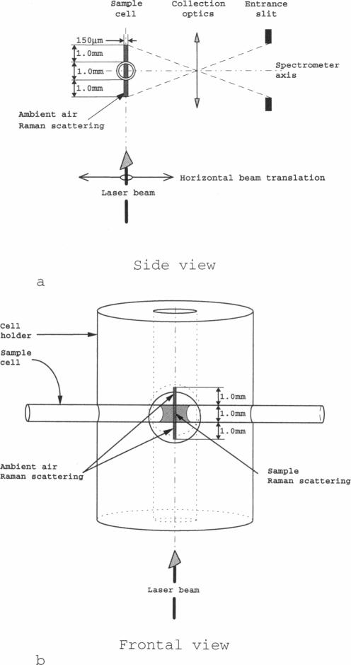
The bond stretching vibration of the cysteine sulfhydryl (SH) group in a typical protein generates a Raman band in the spectral interval 2500-2600 cm-1, a region devoid of interference from any other fundamental mode of vibration of the protein. The relatively high Raman cross section associated with the S-H stretching vibration, the sensitivity of the vibrational frequency to hydrogen bonding interactions and side chain configurations, and the dependence of the Raman intensity on thiol-thiolate equilibria, combine to make the Raman SH band a potentially valuable marker of protein sulfhydryl interactions and a unique indicator of sulfhydryl participation in thiol-disulfide oxidoreductase activity. In order to exploit Raman spectroscopy for these purposes, accurate and precise measurements of Raman SH band profiles are required. We show here that the required precision and accuracy can be achieved by use of the Raman band corresponding to the stretching vibration of in situ nitrogen gas as a quantitative intensity and frequency standard. The Raman Q-branch center of the N2 band occurs at 2330.7 cm-1.

摘要

典型蛋白质中半胱氨酸巯基(SH)基团的键伸缩振动在2500 - 2600 cm-1光谱区间产生拉曼带,该区域没有蛋白质任何其他基本振动模式的干扰。与S - H伸缩振动相关的相对较高的拉曼截面、振动频率对氢键相互作用和侧链构型的敏感性以及拉曼强度对硫醇 - 硫醇盐平衡的依赖性,共同使得拉曼SH带成为蛋白质巯基相互作用的潜在有价值标记以及巯基参与硫醇 - 二硫键氧化还原酶活性的独特指标。为了将拉曼光谱用于这些目的,需要对拉曼SH带轮廓进行准确和精确的测量。我们在此表明,通过使用对应于原位氮气伸缩振动的拉曼带作为定量强度和频率标准,可以实现所需的精度和准确性。N2带的拉曼Q支中心出现在2330.7 cm-1处。

https://cdn.ncbi.nlm.nih.gov/pmc/blobs/b117/1225823/8cef4d707a9d/biophysj00084-0085-a.jpg

相似文献

1
Cysteine conformation and sulfhydryl interactions in proteins and viruses. 3. Quantitative measurement of the Raman S-H band intensity and frequency.蛋白质和病毒中的半胱氨酸构象与巯基相互作用。3. 拉曼S-H带强度和频率的定量测量。
Biophys J. 1993 Sep;65(3):1066-72. doi: 10.1016/S0006-3495(93)81172-X.
2
Distinct cysteine sulfhydryl environments detected by analysis of Raman S-hh markers of Cys-->Ser mutant proteins.通过对半胱氨酸突变为丝氨酸的突变蛋白的拉曼S-hh标记进行分析检测到不同的半胱氨酸巯基环境。
J Mol Biol. 2001 Mar 30;307(3):899-911. doi: 10.1006/jmbi.2001.4476.
3
Structural roles of subunit cysteines in the folding and assembly of the DNA packaging machine (portal) of bacteriophage P22.噬菌体P22的DNA包装机器(门户蛋白)亚基半胱氨酸在折叠和组装中的结构作用。
Biochemistry. 2003 Apr 1;42(12):3437-45. doi: 10.1021/bi020678m.
4
New structural insights from Raman spectroscopy of proteins and their assemblies.蛋白质及其组装体拉曼光谱的新结构见解。
Biopolymers. 2002;67(4-5):214-25. doi: 10.1002/bip.10105.
5
Conformation, stability, and active-site cysteine titrations of Escherichia coli D26A thioredoxin probed by Raman spectroscopy.用拉曼光谱法探测大肠杆菌D26A硫氧还蛋白的构象、稳定性及活性位点半胱氨酸滴定
Protein Sci. 1998 Jan;7(1):193-200. doi: 10.1002/pro.5560070120.
6
Phase transitions in the crystals of L- and DL-cysteine on cooling: intermolecular hydrogen bonds distortions and the side-chain motions of thiol-groups. 1. L-cysteine.L-和DL-半胱氨酸晶体冷却过程中的相变:分子间氢键畸变与硫醇基团的侧链运动。1. L-半胱氨酸
J Phys Chem B. 2008 Oct 9;112(40):12827-39. doi: 10.1021/jp804142c. Epub 2008 Sep 13.
7
Determination of the pKa values of active-center cysteines, cysteines-32 and -35, in Escherichia coli thioredoxin by Raman spectroscopy.通过拉曼光谱法测定大肠杆菌硫氧还蛋白中活性中心半胱氨酸(半胱氨酸-32和-35)的pKa值。
Biochemistry. 1993 Jun 8;32(22):5800-8. doi: 10.1021/bi00073a012.
8
Structure of bacteriophage P22 portal protein in relation to assembly: investigation by Raman spectroscopy.噬菌体P22门户蛋白与组装相关的结构:拉曼光谱研究
Biochemistry. 2001 Nov 13;40(45):13583-91. doi: 10.1021/bi0110488.
9
Absorption and Raman spectroscopy study of cyt c-thiol complexes in acidic solutions.酸性溶液中细胞色素c-硫醇配合物的吸收光谱和拉曼光谱研究
Gen Physiol Biophys. 1992 Jun;11(3):273-86.
10
Weak hydrogen bonds formed by thiol groups in N-acetyl-(L)-cysteine and their response to the crystal structure distortion on increasing pressure.由 N-乙酰-(L)-半胱氨酸中的巯基形成的弱氢键及其对加压下晶体结构变形的响应。
J Phys Chem B. 2013 Nov 21;117(46):14247-60. doi: 10.1021/jp4068872. Epub 2013 Nov 11.

引用本文的文献

1
Large-area high-throughput synthesis of monolayer graphene sheet by Hot Filament Thermal Chemical Vapor Deposition.热丝热化学气相沉积法大面积高通量合成单层石墨烯片。
Sci Rep. 2012;2:682. doi: 10.1038/srep00682. Epub 2012 Sep 21.
2
Conformation, stability, and active-site cysteine titrations of Escherichia coli D26A thioredoxin probed by Raman spectroscopy.用拉曼光谱法探测大肠杆菌D26A硫氧还蛋白的构象、稳定性及活性位点半胱氨酸滴定
Protein Sci. 1998 Jan;7(1):193-200. doi: 10.1002/pro.5560070120.
3
Design and performance of an ultraviolet resonance Raman spectrometer for proteins and nucleic acids.

本文引用的文献

1
ENZYMATIC SYNTHESIS OF DEOXYRIBONUCLEOTIDES. IV. ISOLATION AND CHARACTERIZATION OF THIOREDOXIN, THE HYDROGEN DONOR FROM ESCHERICHIA COLI B.脱氧核糖核苷酸的酶促合成。IV. 来自大肠杆菌B株的氢供体硫氧还蛋白的分离与鉴定
J Biol Chem. 1964 Oct;239:3436-44.
2
Subunit conformational changes accompanying bacteriophage P22 capsid maturation.伴随噬菌体P22衣壳成熟的亚基构象变化。
Biochemistry. 1993 Jan 19;32(2):537-43. doi: 10.1021/bi00053a019.
3
RNA-protein interactions and secondary structures of cowpea chlorotic mottle virus for in vitro assembly.
用于蛋白质和核酸的紫外共振拉曼光谱仪的设计与性能
Biophys J. 1995 Apr;68(4):1607-12. doi: 10.1016/S0006-3495(95)80334-6.
用于体外组装的豇豆花叶病毒的RNA-蛋白质相互作用及二级结构
Biochemistry. 1984 Sep 11;23(19):4301-8. doi: 10.1021/bi00314a008.
4
Thioredoxin and glutaredoxin systems.硫氧还蛋白和谷氧还蛋白系统。
J Biol Chem. 1989 Aug 25;264(24):13963-6.
5
Three-dimensional solution structure of the reduced form of Escherichia coli thioredoxin determined by nuclear magnetic resonance spectroscopy.通过核磁共振光谱法测定的大肠杆菌硫氧还蛋白还原形式的三维溶液结构。
Biochemistry. 1990 May 1;29(17):4129-36. doi: 10.1021/bi00469a016.
6
Crystal structure of thioredoxin from Escherichia coli at 1.68 A resolution.大肠杆菌硫氧还蛋白在1.68埃分辨率下的晶体结构。
J Mol Biol. 1990 Mar 5;212(1):167-84. doi: 10.1016/0022-2836(90)90313-B.
7
Raman spectroscopy of filamentous bacteriophage Ff (fd, M13, f1) incorporating specifically-deuterated alanine and tryptophan side chains. Assignments and structural interpretation.丝状噬菌体Ff(fd、M13、f1)结合特定氘代丙氨酸和色氨酸侧链的拉曼光谱。归属与结构解释。
Biophys J. 1991 Dec;60(6):1337-49. doi: 10.1016/S0006-3495(91)82171-3.
8
Secondary structure and interactions of the packaged dsDNA genome of bacteriophage P22 investigated by Raman difference spectroscopy.通过拉曼差光谱法研究噬菌体P22包装的双链DNA基因组的二级结构和相互作用。
Biochemistry. 1992 Dec 1;31(47):11835-42. doi: 10.1021/bi00162a023.