• 文献检索
  • 文档翻译
  • 深度研究
  • 学术资讯
  • Suppr Zotero 插件Zotero 插件
  • 邀请有礼
  • 套餐&价格
  • 历史记录
应用&插件
Suppr Zotero 插件Zotero 插件浏览器插件Mac 客户端Windows 客户端微信小程序
定价
高级版会员购买积分包购买API积分包
服务
文献检索文档翻译深度研究API 文档MCP 服务
关于我们
关于 Suppr公司介绍联系我们用户协议隐私条款
关注我们

Suppr 超能文献

核心技术专利:CN118964589B侵权必究
粤ICP备2023148730 号-1Suppr @ 2026

文献检索

告别复杂PubMed语法,用中文像聊天一样搜索,搜遍4000万医学文献。AI智能推荐,让科研检索更轻松。

立即免费搜索

文件翻译

保留排版,准确专业,支持PDF/Word/PPT等文件格式,支持 12+语言互译。

免费翻译文档

深度研究

AI帮你快速写综述,25分钟生成高质量综述,智能提取关键信息,辅助科研写作。

立即免费体验

侧脑室中的脑室内巨噬细胞,特别提及室管膜上细胞:定量分析及其对不同年龄大鼠腹腔注射荧光示踪剂的摄取情况

Intraventricular macrophages in the lateral ventricles with special reference to epiplexus cells: a quantitative analysis and their uptake of fluorescent tracer injected intraperitoneally in rats of different ages.

作者信息

Lu J, Kaur C, Ling E A

机构信息

Department of Anatomy, National University of Singapore.

出版信息

J Anat. 1993 Oct;183 ( Pt 2)(Pt 2):405-14.

PMID:8300424
原文链接:https://pmc.ncbi.nlm.nih.gov/articles/PMC1259916/
Abstract

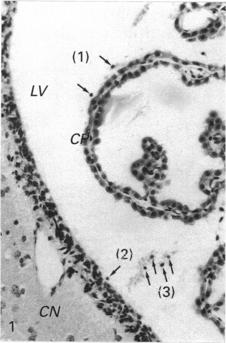
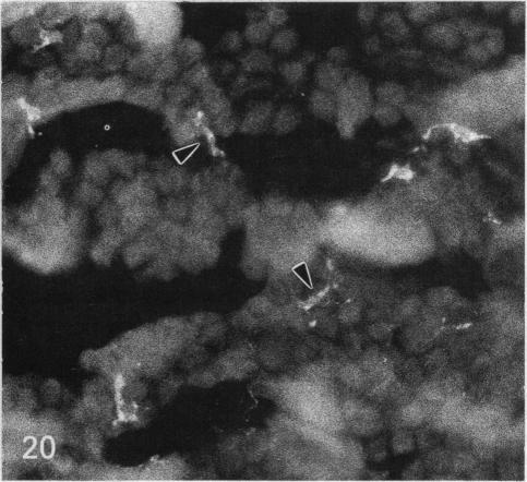
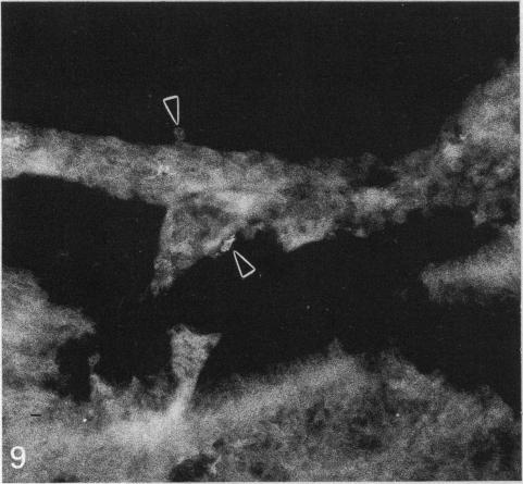
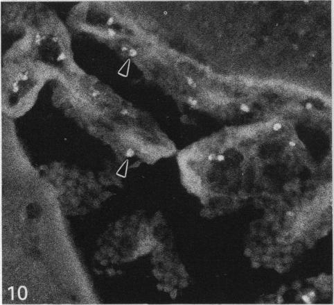
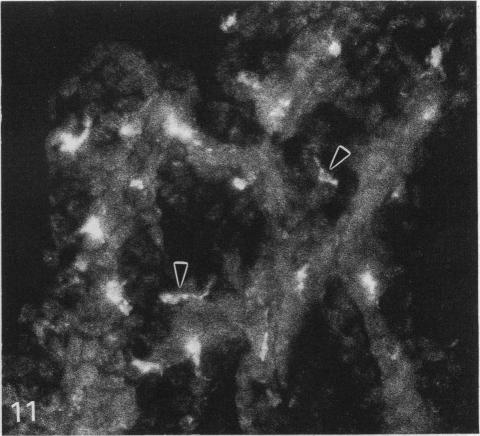
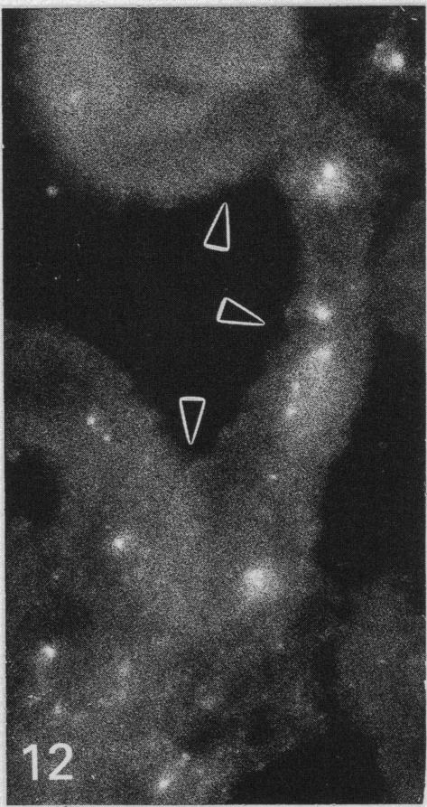
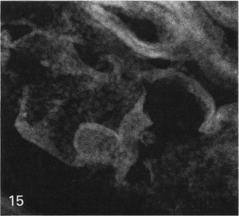
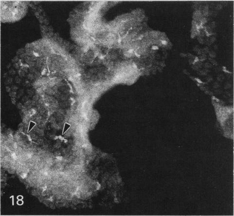
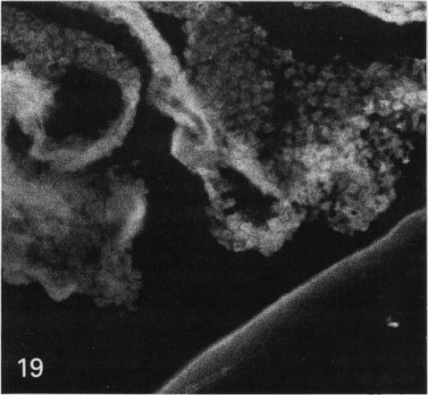
The labelling of epiplexus cells associated with the choroid plexus in the lateral ventricles was examined in rats of different ages with the fluorescent dye, rhodamine isothiocyanate (RhIc). A quantitative study was also attempted; this showed that the number of epiplexus cells and their related cells, namely supraependymal and free-floating cells, increased with age. The mean absolute number of epiplexus cells ranged from approximately 700 in the newborn to approximately 2200 in rats of 17 d of age; thereafter it remained unchanged. The number of free-floating cells also increased substantially but showed considerable individual variation. Following i.p. injection, the tracer was rapidly taken up by the epiplexus cells. This provided strong support for their phagocytic nature. In the newborn (1 d) and developing (13 d, 17 d) rats, RhIc-labelled epiplexus cells were first observed 3 h after the injection. In adult rats, labelled cells were not observed until 12 h after injection. In either case, the fluorescence in the epiplexus cells gradually increased with time. It is suggested from this study that the blood-CSF barrier in the choroid plexus in postnatal rats is incomplete, thereby allowing a rapid transvascular diffusion of the injected RhIc into the blood circulation. The fluorescent dye which enters the ventricle by way of the choroid epithelium is subsequently taken up by the epiplexus cells. Such an unimpeded passage, however, is reduced in the adult rats, probably due to the maturation of the blood capillaries as well as the choroid epithelium.

摘要

用异硫氰酸罗丹明(RhIc)这种荧光染料对不同年龄大鼠侧脑室脉络丛相关的室管膜上细胞进行标记检测。同时还尝试进行了定量研究;结果表明,室管膜上细胞及其相关细胞,即室管膜上和游离漂浮细胞的数量随年龄增加。室管膜上细胞的平均绝对数量范围从新生大鼠的约700个到17日龄大鼠的约2200个;此后保持不变。游离漂浮细胞的数量也大幅增加,但个体差异较大。腹腔注射后,示踪剂迅速被室管膜上细胞摄取。这有力地支持了它们的吞噬性质。在新生(1日龄)和发育中(13日龄、17日龄)大鼠中,注射后3小时首次观察到RhIc标记的室管膜上细胞。在成年大鼠中,直到注射后12小时才观察到标记细胞。无论哪种情况,室管膜上细胞的荧光都随时间逐渐增加。这项研究表明,新生大鼠脉络丛中的血脑脊液屏障不完整,从而使注射的RhIc能迅速经血管扩散到血液循环中。通过脉络丛上皮进入脑室的荧光染料随后被室管膜上细胞摄取。然而,在成年大鼠中,这种畅通无阻的通道减少,可能是由于毛细血管以及脉络丛上皮的成熟。

https://cdn.ncbi.nlm.nih.gov/pmc/blobs/6983/1259916/792b5eaaa6ee/janat00142-0205-f.jpg
https://cdn.ncbi.nlm.nih.gov/pmc/blobs/6983/1259916/94bcfabe02d3/janat00142-0199-a.jpg
https://cdn.ncbi.nlm.nih.gov/pmc/blobs/6983/1259916/996535bdd4a0/janat00142-0199-b.jpg
https://cdn.ncbi.nlm.nih.gov/pmc/blobs/6983/1259916/033d9fce0a8e/janat00142-0199-c.jpg
https://cdn.ncbi.nlm.nih.gov/pmc/blobs/6983/1259916/ee497c6c70b1/janat00142-0199-d.jpg
https://cdn.ncbi.nlm.nih.gov/pmc/blobs/6983/1259916/f5442b4e256a/janat00142-0203-a.jpg
https://cdn.ncbi.nlm.nih.gov/pmc/blobs/6983/1259916/0e510e88fc29/janat00142-0203-b.jpg
https://cdn.ncbi.nlm.nih.gov/pmc/blobs/6983/1259916/fbb72975ab07/janat00142-0203-c.jpg
https://cdn.ncbi.nlm.nih.gov/pmc/blobs/6983/1259916/382505706dca/janat00142-0203-d.jpg
https://cdn.ncbi.nlm.nih.gov/pmc/blobs/6983/1259916/8e7fb781621d/janat00142-0203-e.jpg
https://cdn.ncbi.nlm.nih.gov/pmc/blobs/6983/1259916/a2ec449b6e4f/janat00142-0203-f.jpg
https://cdn.ncbi.nlm.nih.gov/pmc/blobs/6983/1259916/3f15a8212a1b/janat00142-0203-g.jpg
https://cdn.ncbi.nlm.nih.gov/pmc/blobs/6983/1259916/96cd2daaa521/janat00142-0205-a.jpg
https://cdn.ncbi.nlm.nih.gov/pmc/blobs/6983/1259916/312edb2d1fe2/janat00142-0205-b.jpg
https://cdn.ncbi.nlm.nih.gov/pmc/blobs/6983/1259916/2d4d7ae66416/janat00142-0205-c.jpg
https://cdn.ncbi.nlm.nih.gov/pmc/blobs/6983/1259916/a935bb5f0f1a/janat00142-0205-d.jpg
https://cdn.ncbi.nlm.nih.gov/pmc/blobs/6983/1259916/95fc8483de7d/janat00142-0205-e.jpg
https://cdn.ncbi.nlm.nih.gov/pmc/blobs/6983/1259916/792b5eaaa6ee/janat00142-0205-f.jpg

相似文献

1
Intraventricular macrophages in the lateral ventricles with special reference to epiplexus cells: a quantitative analysis and their uptake of fluorescent tracer injected intraperitoneally in rats of different ages.侧脑室中的脑室内巨噬细胞,特别提及室管膜上细胞:定量分析及其对不同年龄大鼠腹腔注射荧光示踪剂的摄取情况
J Anat. 1993 Oct;183 ( Pt 2)(Pt 2):405-14.
2
Origin, nature, and some functional considerations of intraventricular macrophages, with special reference to the epiplexus cells.脑室内巨噬细胞的起源、性质及一些功能方面的考量,特别提及脉络丛细胞。
Microsc Res Tech. 1998 Apr 1;41(1):43-56. doi: 10.1002/(SICI)1097-0029(19980401)41:1<43::AID-JEMT5>3.0.CO;2-V.
3
Effects of dexamethasone on the epiplexus cells in postnatal rats.地塞米松对新生大鼠脉络丛细胞的影响。
Neurosci Lett. 1995 Aug 25;196(3):165-8. doi: 10.1016/0304-3940(95)11866-u.
4
Uptake of tracer by the epiplexus cells via the choroid plexus epithelium following an intravenous or intraperitoneal injection of horseradish peroxidase in rats.在大鼠静脉内或腹腔内注射辣根过氧化物酶后,示踪剂通过脉络丛上皮被室管膜上细胞摄取。
J Anat. 1993 Dec;183 ( Pt 3)(Pt 3):609-17.
5
Expression and upregulation of transferrin receptors and iron uptake in the epiplexus cells of different aged rats injected with lipopolysaccharide and interferon-gamma.注射脂多糖和γ干扰素的不同年龄大鼠室管膜上细胞中转铁蛋白受体的表达、上调及铁摄取
J Anat. 1995 Dec;187 ( Pt 3)(Pt 3):603-11.
6
Labelling of retinal microglial cells following an intravenous injection of a fluorescent dye into rats of different ages.将荧光染料静脉注射到不同年龄的大鼠体内后视网膜小胶质细胞的标记
J Anat. 2000 Feb;196 ( Pt 2)(Pt 2):173-9. doi: 10.1046/j.1469-7580.2000.19620173.x.
7
Variation with age in the labelling of amoeboid microglial cells in rats following intraperitoneal or intravenous injection of a fluorescent dye.腹腔内或静脉内注射荧光染料后,大鼠中阿米巴样小胶质细胞标记随年龄的变化。
J Anat. 1993 Feb;182 ( Pt 1)(Pt 1):55-63.
8
Effects of chloroquine on the ependyma, choroid plexus and epiplexus cells in the lateral ventricles of rats.氯喹对大鼠侧脑室室管膜、脉络丛和室管膜上细胞的影响。
J Hirnforsch. 1997;38(1):99-106.
9
Immunophenotypic features of epiplexus cells and their response to interferon gamma injected intraperitoneally in postnatal rats.产后大鼠脉络丛细胞的免疫表型特征及其对腹腔注射干扰素γ的反应。
J Anat. 1994 Aug;185 ( Pt 1)(Pt 1):75-84.
10
[The development of epiplexus cells (Kolmer cells) in the choroid plexus of the fourth ventricle of the mouse. A scanning and transmission electron microscopic study].[小鼠第四脑室脉络丛中室管膜周细胞(科尔默细胞)的发育。扫描和透射电子显微镜研究]
Z Mikrosk Anat Forsch. 1980;94(2):316-26.

引用本文的文献

1
Role of macrophages in pulmonary arterial hypertension.巨噬细胞在肺动脉高压中的作用。
Front Immunol. 2023 Apr 19;14:1152881. doi: 10.3389/fimmu.2023.1152881. eCollection 2023.
2
The choroid plexus and its role in the pathogenesis of neurological infections.脉络丛及其在神经感染发病机制中的作用。
Fluids Barriers CNS. 2022 Sep 10;19(1):75. doi: 10.1186/s12987-022-00372-6.
3
Transmigration of macrophages across the choroid plexus epithelium in response to the feline immunodeficiency virus.巨噬细胞穿越脉络丛上皮细胞的转位反应与猫免疫缺陷病毒有关。

本文引用的文献

1
The electron microscopy of the choroid plexus.脉络丛的电子显微镜检查。
J Biophys Biochem Cytol. 1956 Jul 25;2(4):467-74. doi: 10.1083/jcb.2.4.467.
2
[Experimental study of the function and origin of Kolmer's cells of plexus chorioideus in axolotl and guinea pig].[美西螈和豚鼠脉络丛科尔默细胞的功能与起源的实验研究]
Z Anat Entwicklungsgesch. 1953;117(1):1-19.
3
Supraependymal macrophages of third ventricle of hamster: morphological, functional and histochemical characterization in situ and in culture.仓鼠第三脑室室管膜上巨噬细胞:原位及培养状态下的形态学、功能及组织化学特征
Cell Tissue Res. 2012 Feb;347(2):443-55. doi: 10.1007/s00441-011-1301-8. Epub 2012 Jan 27.
4
Choroid plexus macrophages proliferate and release toxic factors in response to feline immunodeficiency virus.脉络丛巨噬细胞会因猫免疫缺陷病毒而增殖并释放毒性因子。
J Neurovirol. 2002 Jun;8(3):225-39. doi: 10.1080/13550280290049679.
5
Morphological studies of the hyalocytes in the chicken eye: scanning electron microscopy and inflammatory response after the intravitreous injection of carbon particles.鸡眼中透明细胞的形态学研究:玻璃体腔内注射碳颗粒后的扫描电子显微镜观察及炎症反应
J Anat. 1996 Jun;188 ( Pt 3)(Pt 3):661-9.
6
An immunohistochemical study of the intraventricular macrophages in induced hydrocephalus in prenatal rats following a maternal injection of 6-aminonicotinamide.对孕鼠注射6-氨基烟酰胺后诱导的产前大鼠脑积水模型脑室内巨噬细胞的免疫组织化学研究。
J Anat. 1996 Apr;188 ( Pt 2)(Pt 2):491-5.
7
Expression and upregulation of transferrin receptors and iron uptake in the epiplexus cells of different aged rats injected with lipopolysaccharide and interferon-gamma.注射脂多糖和γ干扰素的不同年龄大鼠室管膜上细胞中转铁蛋白受体的表达、上调及铁摄取
J Anat. 1995 Dec;187 ( Pt 3)(Pt 3):603-11.
8
Uptake of tracer by the epiplexus cells via the choroid plexus epithelium following an intravenous or intraperitoneal injection of horseradish peroxidase in rats.在大鼠静脉内或腹腔内注射辣根过氧化物酶后,示踪剂通过脉络丛上皮被室管膜上细胞摄取。
J Anat. 1993 Dec;183 ( Pt 3)(Pt 3):609-17.
9
Immunophenotypic features of epiplexus cells and their response to interferon gamma injected intraperitoneally in postnatal rats.产后大鼠脉络丛细胞的免疫表型特征及其对腹腔注射干扰素γ的反应。
J Anat. 1994 Aug;185 ( Pt 1)(Pt 1):75-84.
J Comp Neurol. 1980 Aug 1;192(3):489-504. doi: 10.1002/cne.901920308.
4
Ultrastructure and mode of formation of epiplexus cells in the choroid plexus in the lateral ventricles of the monkey (Macaca fascicularis).猕猴(食蟹猴)侧脑室脉络丛中室管膜上细胞的超微结构及形成方式
J Anat. 1981 Dec;133(Pt 4):555-69.
5
Light and electron microscopic demonstration of non-specific esterase in amoeboid microglial cells in the corpus callosum in postnatal rats: a cytochemical link to monocytes.出生后大鼠胼胝体中阿米巴样小胶质细胞非特异性酯酶的光镜和电镜显示:与单核细胞的细胞化学联系
J Anat. 1982 Sep;135(Pt 2):385-94.
6
A light microscopic and scanning electron microscopic study of intraventricular macrophages in the brains of aged mice.老年小鼠脑室内巨噬细胞的光镜和扫描电镜研究
J Anat. 1983 Jun;136(Pt 4):761-71.
7
Scanning electron microscopic study of epiplexus cells in the lateral ventricles of the monkey (Macaca fascicularis).猕猴(食蟹猴)侧脑室脉络丛细胞的扫描电子显微镜研究。
J Anat. 1983 Dec;137 ( Pt 4)(Pt 4):645-52.
8
Structural aspects of brain barriers, with special reference to the permeability of the cerebral endothelium and choroidal epithelium.脑屏障的结构方面,特别提及脑内皮和脉络膜上皮的通透性。
Int Rev Cytol. 1980;65:117-91. doi: 10.1016/s0074-7696(08)61960-9.
9
Electron microscopic study of the epiplexus (Kolmer) cells of the cat choroid plexus.猫脉络丛室管膜下(科尔默)细胞的电子显微镜研究。
Z Zellforsch Mikrosk Anat. 1970;110(4):471-86. doi: 10.1007/BF00330099.
10
Scanning electron microscope observation of intraventricular macrophages (Kolmer cells) in the rat brain.大鼠脑室内巨噬细胞(科尔默细胞)的扫描电子显微镜观察
Arch Histol Jpn. 1973 Jan;35(2):133-40. doi: 10.1679/aohc1950.35.133.