Müller J G, Krenn V, Schindler C, Czub S, Stahl-Hennig C, Coulibaly C, Hunsmann G, Kneitz C, Kerkau T, Rethwilm A
Institute of Pathology, University of Würzburg, Germany.
Am J Pathol. 1993 Sep;143(3):699-713.
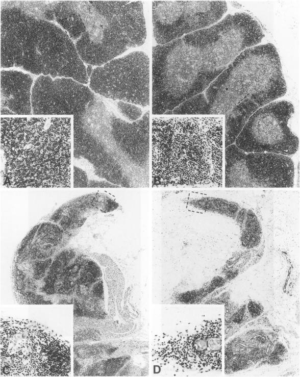
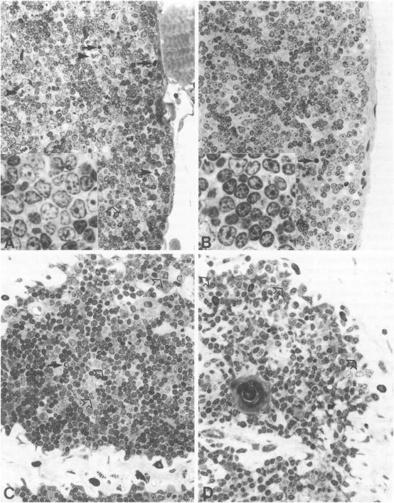
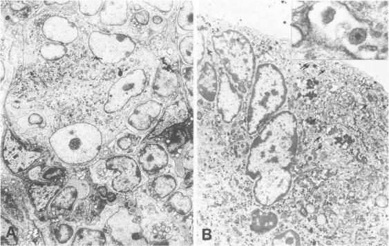
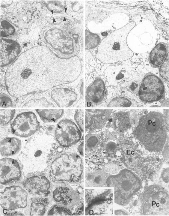
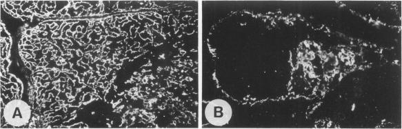
The role of the thymus in the pathogenesis of simian acquired immunodeficiency syndrome was investigated in 18 juvenile rhesus monkeys (Macaca mulatta). The thymus was infected from the first week post-SIVmac inoculation, but the amount of virus-positive cells was very low (< 1 in 10(4) T cells) as demonstrated by polymerase chain reaction and in situ hybridization. First morphological alteration was a narrowing of the cortex at 12 and 24 wpi. Morphometry revealed no increase of pyknotic T cells but a decrease of the proliferation rate and flow cytometry showed a reduction of the immature CD4+/CD8+ double-positive T cells. Ultrastructural analysis revealed vacuolization, shrinkage, and finally cytolysis of the cortical epithelial cells and the interdigitating dendritic cells. Immunofluorescence staining exhibited a widespread loss of cortical epithelial cells. This damage to the thymic microenvironment could explain the breakdown of the intrathymic T cell proliferation. It preceded fully developed simian acquired immunodeficiency syndrome and is therefore considered to play a major role in its pathogenesis.
在18只幼年恒河猴(猕猴)中研究了胸腺在猴获得性免疫缺陷综合征发病机制中的作用。从接种SIVmac后的第一周开始,胸腺就受到感染,但通过聚合酶链反应和原位杂交证明,病毒阳性细胞的数量非常少(每10⁴个T细胞中少于1个)。最初的形态学改变是在接种后12周和24周时皮质变窄。形态计量学显示固缩性T细胞没有增加,但增殖率降低,流式细胞术显示未成熟的CD4⁺/CD8⁺双阳性T细胞减少。超微结构分析显示皮质上皮细胞和交错突细胞出现空泡化、萎缩,最终细胞溶解。免疫荧光染色显示皮质上皮细胞广泛丢失。胸腺微环境的这种损伤可以解释胸腺内T细胞增殖的破坏。它先于完全发展的猴获得性免疫缺陷综合征出现,因此被认为在其发病机制中起主要作用。