Leforestier A, Livolant F
Centre de Biologie Cellulaire, Ivry-sur-Seine, France.
Biophys J. 1993 Jul;65(1):56-72. doi: 10.1016/S0006-3495(93)81063-4.
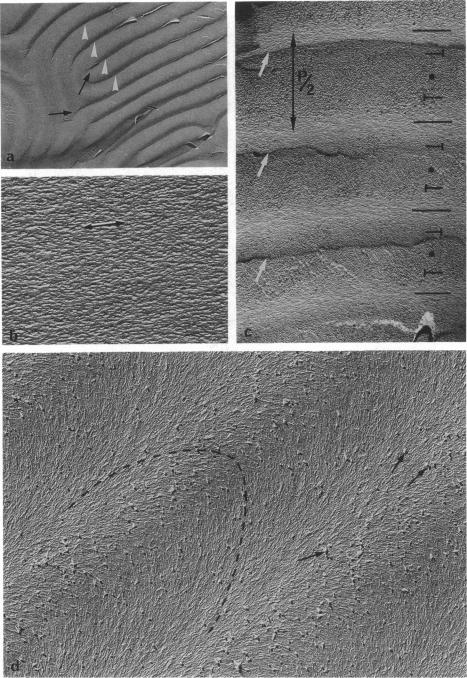
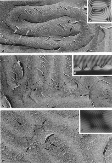
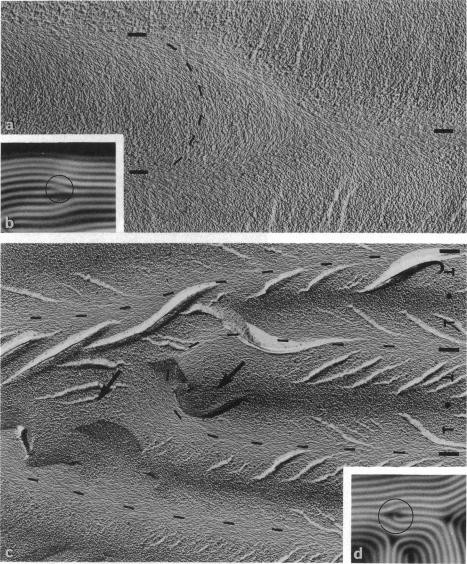
Aqueous solutions of 146-base pair DNA fragments form a cholesteric liquid crystalline phase in the range of about 160-290 mg/ml. We present a structural analysis of this phase by comparing the data obtained from polarizing and electron microscopy. This phase shows multiple aspects or "textures" which are presented and interpreted. They mainly depend on the orientation of the structure relative to the observation plane and on the nature, distribution, and amount of defects present in the phase. These defects are then analyzed with the two methods, and the molecular orientations can be defined precisely in their core. The biological interest of such structural analyses is discussed in relation with the understanding of chromatin structure and function.
146个碱基对的DNA片段的水溶液在约160 - 290毫克/毫升的范围内形成胆甾相液晶相。我们通过比较偏振显微镜和电子显微镜获得的数据,对该相进行了结构分析。该相呈现出多种特征或“纹理”,并对其进行了展示和解释。它们主要取决于结构相对于观察平面的取向,以及该相中存在的缺陷的性质、分布和数量。然后用这两种方法对这些缺陷进行分析,并能在其核心精确界定分子取向。结合对染色质结构和功能的理解,讨论了这种结构分析的生物学意义。