Aikawa M, Komata Y, Asai T, Midorikawa O
Am J Pathol. 1977 May;87(2):285-96.
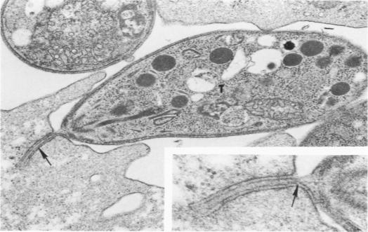
Entry by tachyzoites of Toxoplasma gondii, the causative agent of toxoplasmosis, into peritoneal cells was investigated with transmission and scanning electron microscopy. The process of entry is initiated by the parasite contacting the host cell with its anterior end, creating a small depression in the plasmalemma of the host cell. Occasionally, a small portion of the host cell cytoplasm protrudes and contaccts the anterior end of the parasite. A cylindrical structure (35 nm in diameter) extends from the pellicle of the parasite to the host cell. Such structures appear to assist host cell entry by T. gondii. As the entry process progresses, pseudopods of the host cell surround theparasite and finally T gondii becomes intracellular, being located in a vacuole separated from the host cell cytoplasm by a unit membrane. (Am J. Pathol 87:285-296, 1977).
利用透射电子显微镜和扫描电子显微镜研究了弓形虫病的病原体刚地弓形虫速殖子进入腹膜细胞的过程。进入过程始于寄生虫前端与宿主细胞接触,在宿主细胞质膜上形成一个小凹陷。偶尔,宿主细胞的一小部分细胞质会突出并接触到寄生虫的前端。一种圆柱形结构(直径35纳米)从寄生虫的表膜延伸到宿主细胞。这些结构似乎有助于刚地弓形虫进入宿主细胞。随着进入过程的推进,宿主细胞的伪足包围寄生虫,最终刚地弓形虫进入细胞内,位于一个由单位膜与宿主细胞质隔开的液泡中。(《美国病理学杂志》87:285 - 296, 1977年)